Iatrogenic Subclavian Artery Perforation Rescued by Operator-Modified Graft Stent
- Affiliations
-
- 1Divisions of Interventional Cardiology and Adult Cardiac Surgery, Cardiovascular Center, Taichung Veterans General Hospital, Taichung, Taiwan, R.O.C. wenliengleevghtc@gmail.com
- 2School of Medicine, National Yang-Ming University, Taipei, Taiwan, R.O.C.
- KMID: 2427139
- DOI: http://doi.org/10.3349/ymj.2017.58.2.462
Abstract
- Subclavian artery (SCA) perforation is a rare complication while performing SCA intervention. In our present report, a 73-year-old female, with stenosis of the left SCA and situs inversus, presented with exercise-induced left arm weakness. The SCA stenosis was treated with direct stenting with a balloon-expansible Express LD 10×25 mm stent. However, it caused iatrogenic SCA perforation and hemothorax. The perforation was sealed by endovascular repair with operator-modified Endurant II graft stent, which complicated with occlusion of left common carotid artery. And, the carotid artery was rescued by another stent. The graft stent, which was originally designed for abdominal aortic aneurysm, can be modified to suitable length and take as a rescue stent of large vessel with iatrogenic perforation. Due to strong radial force of graft stent, preservation of large side branches should been watched out.
Keyword
MeSH Terms
Figure
-
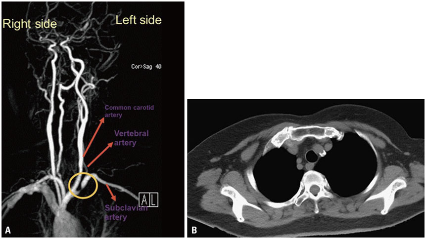
Fig. 1 Magnetic resonance angiogram (MRA) and computer tomography (CT) of the upper limb vessels. (A) MRA shows severe stenosis in the proximal left subclavian artery (circle). (B) CT reveals calcification near the orifice of left subclavian artery.
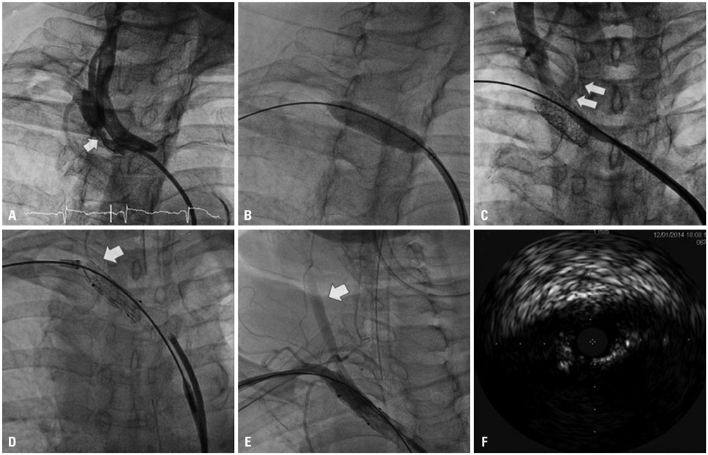
Fig. 2 Angiography with anterior view of the left brachiocephaic trunk and left subclavian artery (SCA) showing proximal severe stenosis (these images are all mirrored image of this patient with situs inversus; that is, it look-like “right brachiocephaic trunk in images” actually are left ones). (A) Severe stenosis in the proximal SCA (arrow). (B) Direct stenting by an Express LD 10×25 mm stent. (C) SCA perforation with contrast extravasation (arrows). (D) Retrograde approach through the left brachial artery: covered by the graft stent (arrow). (E) Slow flow in the left common carotid artery (arrow) due to ostium occluded by the graft stent. (F) Intravascular ultrasound shows severe occlusion by the graft stent.

Fig. 3 The procedure of modification of graft stent. (A and B) After deployed out graft stent, the stent was cut between ring and ring and to a wanted length. (C) Using sutures to aid collapse of the ring of deployed stent. (D) With the compression of sutures, the stent could be resheathed into sheath. (E) Repeating the same way, the stent was resheathed ring by ring. (F) Resheathed modified stents ready to be deployed.
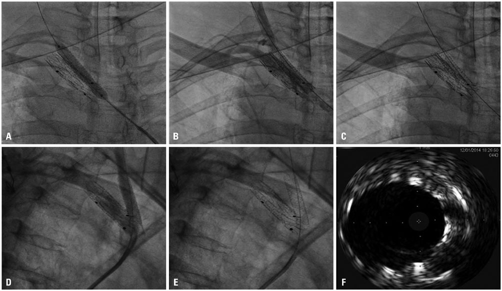
Fig. 4 Angiographic images during rewiring and stenting of the left common carotid artery (LCCA) (these images are all mirrored image of this patient with situs inversus; that is, the “right brachiocephaic trunk in images” actually are left ones). (A) Stenting from the LCCA to the left brachiocephalic artery. (B and C) After stenting, frontal view of angiography after (B) and before (C) contrast injection. (D and E) After stenting, lateral view of angiography after (D) and before (E) contrast injection. (F) Intravascular ultrasound shows the ostium of the LCCA re-opened by the stent.
Reference
-
1. Patel SN, White CJ, Collins TJ, Daniel GA, Jenkins JS, Reilly JP, et al. Catheter-based treatment of the subclavian and innominate arteries. Catheter Cardiovasc Interv. 2008; 71:963–968.
Article2. Ochoa VM, Yeghiazarians Y. Subclavian artery stenosis: a review for the vascular medicine practitioner. Vasc Med. 2011; 16:29–34.
Article3. Takach TJ, Duncan JM, Livesay JJ, Krajcer Z, Cervera RD, Gregoric ID, et al. Brachiocephalic reconstruction II: operative and endovascular management of single-vessel disease. J Vasc Surg. 2005; 42:55–61.
Article
- Full Text Links
-
- Actions
-
Cited
- CITED
-
- Close
- Share
-
- Similar articles
-
- Urgent Endovascular Stent Graft Placement for Iatrogenic Subclavian Artery Rupture
- Endovascular Repair of Thoracic Aortic Aneurysm Using a Custom-made Fenestrated Stent Graft to Preserve the Left Subclavian Artery
- Coronary Artery Perforation Following Implantation of a Drug-Eluting Stent Rescued by Deployment of a Covered Stent in Symptomatic Myocardial Bridging
- Iatrogenic Subclavian Artery Aneurysm: Report of a Case
- A Hybrid Procedure for Coronary Artery Disease with Left Subclavian Artery Stenosis
