J Korean Ophthalmol Soc.
2018 Nov;59(11):1009-1016. 10.3341/jkos.2018.59.11.1009.
Comparative Effect of Spectacles and Orthokeratology Lenses on Axial Elongation in Children with Mild-to-Moderate Myopia
- Affiliations
-
- 1Saevit Eye Hospital, Goyang, Korea. jjulujjulu@hanmail.net
- 2Jalboneun Seoul Eye Clinic, Hwaseoung, Korea.
- KMID: 2426361
- DOI: http://doi.org/10.3341/jkos.2018.59.11.1009
Abstract
- PURPOSE
To assess the effect on axial elongation and associated factors between spectacles and of orthokeratology lens (OK) wearing in children with mild to moderate myopia.
METHODS
A total of one hundred subjects, ranging in age from 6 to 13 years, and with mild to moderate myopia no more than −4.50 diopters in spherical equivalent, visited our clinic from 2013 to 2015. The OK group (75 eyes) and the spectacles group (64 eyes) were compared and analyzed on the axial elongation and associated factors.
RESULTS
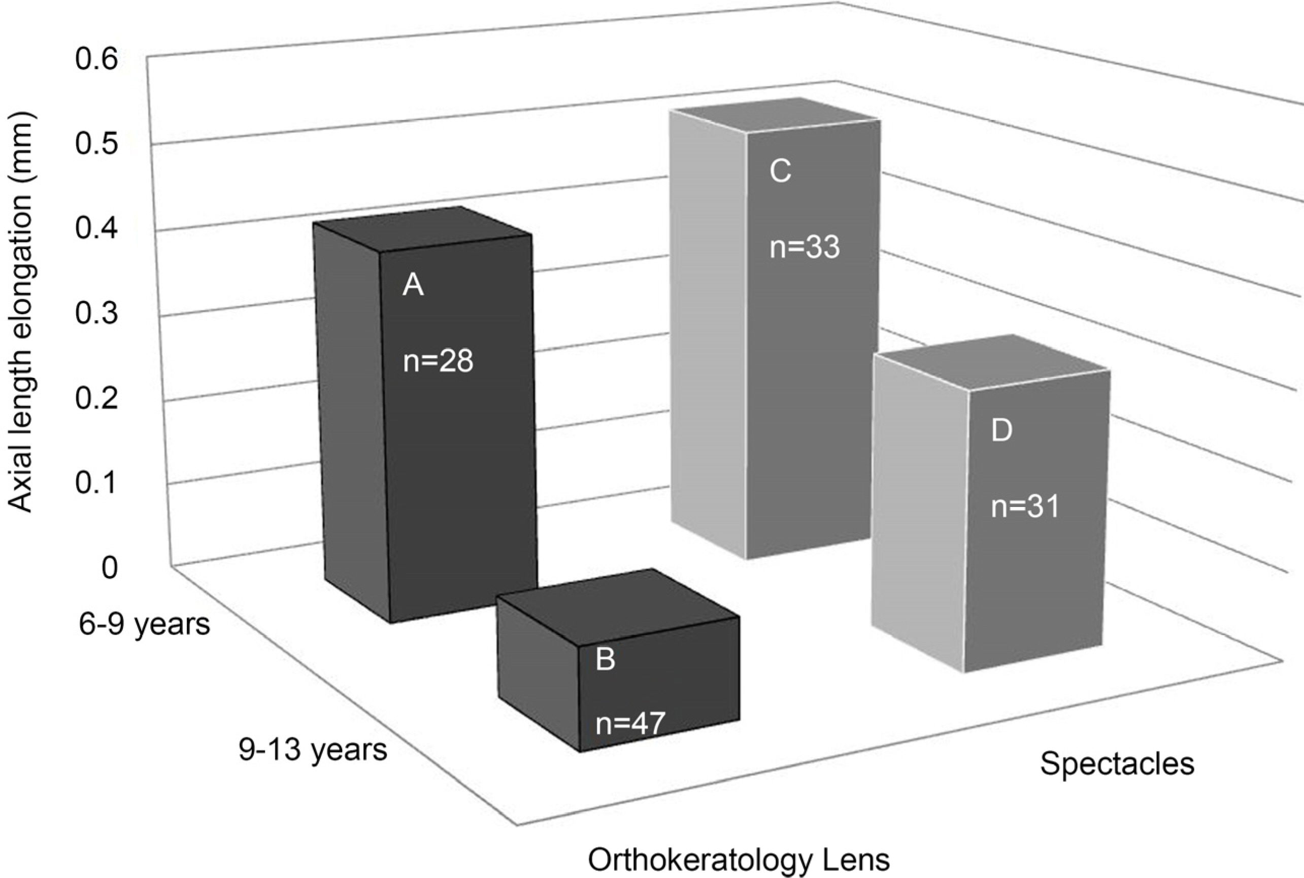
In the OK group, axial length was elongated in 1 year period with a mean increase of 0.24 ± 0.29 mm. In spectacles group, axial length was elongated in 1 year period with a mean increase of 0.42 ± 0.20 mm. The statistically significant suppression of axial elongation was observed in OK group compared to the spectacles group (Mann-Whitney U test, p < 0.05). For OK group, the age of starting OK (Pearson's correlation, r = −0.481, p < 0.05) was the only influencing factor on axial elongation, which had negative correlation with axial elongation. In spectacles group, the age of starting spectacles had negative correlation with axial elongation (Pearson's correlation, r = −0.462, p < 0.05) and baseline spherical equivalent, spherical diopter, cylindrical diopter from manifest refraction had positive correlation with axial elongation. Comparison of axial elongation in orthokeratology lens group and spectacles group by age groups (6 to 9 years [28 eyes], 9 to 13 years [47 eyes]), 9 to 13 years of orthokeratology lens group had the stronger suppression of axial elongation (Mann-Whitney U test, p < 0.05).
CONCLUSIONS
The OK effectively suppresses axial elongation compared to the spectacles. Although the patients are in age from 9 to 13 years, the axial elongation was effectively suppressed.
Keyword
MeSH Terms
Figure
Reference
-
References
1. Holden BA, Fricke TR, Wilson DA, et al. Global prevalence of abdominal and high myopia and temporal trends from 2000 through 2050. Ophthalmology. 2016; 123:1036–42.2. Koh V, Yang A, Saw SM, et al. Differences in prevalence of abdominal errors in young asian males in Singapore between 1996–1997 and 2009–2010. Ophthalmic Epidemiol. 2014; 21:247–55.3. Lee YY, Lo CT, Sheu SJ, Lin JL. What factors are associated with myopia in young adults? A survey study in Taiwan Military Conscripts. Invest Ophthalmol Vis Sci. 2013; 54:1026–33.
Article4. Jung S, Han J, Kwon J, et al. Analysis of myopic progression in childhood using the Korea National Health and Nutrition Examination Survey. J Korean Ophthalmol Soc. 2016; 57:1430–4.
Article5. The Eye Disease Case-Control Study Group. Risk factors for abdominal macular holes. Am J Ophthalmol. 1994; 118:754–61.6. Mitchell P, Hourihan F, Sandbach J, Wang JJ. The relationship abdominal glaucoma and myopia: the Blue Mountains Eye Study. Ophthalmology. 1999; 106:2010–5.7. Saw SM, Gazzard G, Shih– Yen EC, Chua WH. Myopia and abdominal pathological complications. Ophthalmic Physiol Opt. 2005; 25:381–91.8. Harper AR, Summers JA. The dynamic sclera: extracellular matrix remodeling in normal ocular growth and myopia development. Exp Eye Res. 2015; 133:100–11.
Article9. Schwartz JT. Results of a monozygotic cotwin control study on a treatment for myopia. Prog Clin Biol Res. 1981; 69:249–58.10. Yen MY, Liu JH, Kao SC, Shiao CH. Comparison of the effect of atropine and cyclopentolate on myopia. Ann Ophthalmol. 1989; 21:180–2.11. Shih YF, Chen CH, Chou AC, et al. Effects of different abdominal of atropine on controlling myopia in myopic children. J Ocul Pharmacol Ther. 1999; 15:85–90.12. Siatkowski RM, Cotter S, Miller JM, et al. Safety and efficacy of 2% pirenzepine ophthalmic gel in children with myopia: a 1– year, multicenter, double– masked, placebo– controlled parallel study. Arch Ophthalmol. 2004; 122:1667–74.13. Tan DT, Lam DS, Chua WH, et al. One– year multicenter, double– masked, placebo– controlled, parallel safety and efficacy study of 2% pirenzepine ophthalmic gel in children with myopia. Ophthalmology. 2005; 112:84–91.14. Jensen H. Timolol maleate in the control of myopia. A preliminary report. Acta Ophthalmol Suppl (Oxf). 1988; 185:128–9.
Article15. Edwards MH, Li Rw, Lam CS, et al. The Hong Kong progressive lens myopia control study: study design and main findings. Invest Ophthalmol Vis Sci. 2002; 43:2852–8.16. Gwiazda J, Hyman L, Hussein M, et al. A randomized clinical trial of progressive addition lenses versus single vision lenses on the progression of myopia in children. Invest Ophthalmol Vis Sci. 2003; 44:1492–500.
Article17. Walline JJ, Jones LA, Mutti DO, Zadnik K. A randomized trial of the effects of rigid contact lenses on myopia progression. Arch Ophthalmol. 2004; 122:1760–6.18. Walline JJ, Lindsley K, Vedula SS, et al. Interventions to slow abdominal of myopia in children. Cochrane Database Syst Rev. 2011; 12:CD004916. doi:. DOI: 10.1002/14651858.CD004916.19. Nichols JJ, Marsich MM, Nguyen M, et al. Overnight orthokeratology. Optom Vis Sci. 2000; 77:252–9.
Article20. Swarbrick HA, Wong G, O'Leary DJ. Corneal response to orthokeratology. Optom Vis Sci. 1998; 75:791–9.
Article21. Park YM, Lee JH, Park YK, et al. Effect of toric orthokeratology lenses in patients with limbus to limbus corneal astigmatism. J Korean Ophthalmol Soc. 2015; 56:830–4.
Article22. Lee SH, Lee DH, Lee HK. Analysis of the cause of failure in the correction of childhood myopia using orthokeratologic lenses. J Korean Ophthalmol Soc. 2015; 56:317–22.
Article23. Kim JR, Chung TY, Lim DH, Bae JH. Effect of orthokeratologic lenses on myopic progression in childhood. J Korean Ophthalmol Soc. 2013; 54:401–7.
Article24. Lee WH, Park YK, Seo JM, Shin JH. The inhibitory effect of abdominal and astigmatic progression by orthokeratology lens. J Korean Ophthalmol Soc. 2011; 52:1269–74.25. Cheung SW, Cho P, Fan D. Asymmetrical increase in axial length in the two eyes of a monocular orthokeratology patient. Optom Vis Sci. 2004; 81:653–6.
Article26. Cho P, Cheung SW. Retardation of myopia in Orthokeratology (ROMIO) study: a 2– year randomized clinical trial. Invest Ophthalmol Vis Sci. 2012; 53:7077–85.27. Charman WN, Mountford J, Atchison DA, Markwell EL. Peripheral refraction in orthokeratology patients. Optom Vis Sci. 2006; 83:641–8.
Article28. Cho P, Cheung SW, Edwards M. The longitudinal orthokeratology research in children (LORIC) in Hong Kong: a pilot study on abdominal changes and myopic control. Curr Eye Res. 2005; 30:71–80.29. Walline JJ, Jones LA, Sinnott LT. Corneal reshaping and myopia progression. Br J Ophthalmol. 2009; 93:2852–8.
Article30. Kakita T, Hiraoka T, Oshika T. Influence of overnight abdominal on axial elongation in childhood myopia. Invest Ophthalmol Vis Sci. 2011; 52:2170–4.31. Edwards MH. The development of myopia in Hong Kong children between the ages of 7 and 12 years: a five– year longitudinal study. Ophthalmic Physiol Opt. 1999; 19:286–94.32. Jones LA, Mitchell GL, Mutti DO, et al. Comparison of ocular component growth curves among refractive error groups in children. Invest Ophthalmol Vis Sci. 2005; 46:2317–27.
Article33. Hyman L, Gwiazda J, Hussein M, et al. Relationship of age, sex, ethnicity with myopia progression and axial elongation in the abdominal of myopia evaluation trial. Arch Ophthalmol. 2005; 123:977–87.34. Goss DA, Winkler RL. Progression of myopia in youth: age of cessation. Am J Optom Vis Sci. 1983; 83:651–8.35. Hiraoka T, Kakita T, Okamoto F, et al. Long– term effect of abdominal orthokeratology on axial length elongation in childhood abdominal: a 5– year follow– up study. Invest Ophthalmol Vis Sci. 2012; 53:3913–9.36. Charm J, Cho P. High myopia– partial reduction ortho– k: a 2– year randomized study. Optom Vis Sci. 2013; 90:530–9.37. Fu AC, Chen XL, Lv Y, et al. Higher spherical equivalent refractive errors is associated with slower axial elongation wearing orthokeratology. Cont Lens Anterior Eye. 2016; 39:62–6.
Article38. Zhong Y, Chen Z, Xue F, et al. Corneal power change is predictive of myopia progression in orthokeratology. Optom Vis Sci. 2014; 91:404–11.
Article39. Santodomingo– Rubido J, Villa– Collar C, Gilmartin B, et al. Factors preventing myopia progression with orthokeratology correction. Optom Vis Sci. 2013; 90:1225–36.
- Full Text Links
- Actions
-
Cited
- CITED
-
- Close
- Share
- Similar articles
-
- Influence of Orthokeratology Lens on Axial length Elongation and Myopic Progression in Childhood Myopia
- Comparative Analysis of Orthokeratology Lenses and Low-Concentration Atropine Eye Drops on Axial Length Elongation
- Prevention of myopia progression using orthokeratology
- Clinical Characteristics of Prescribing Orthokeratology Lenses in Adult
- Prescription and effect of orthokeratology lenses


