Clin Endosc.
2013 Jul;46(4):414-417.
Endoscopic Treatment of a Symptomatic Ileal Lipoma with Recurrent Ileocolic Intussusceptions by Using Cap-Assisted Colonoscopy
- Affiliations
-
- 1Department of Internal Medicine, Hanyang University College of Medicine, Seoul, Korea. drkangmd@naver.com
Abstract
- A 73-year-old woman presented with intermittent abdominal pain and weight loss of 15 kg for 2 years. Colonoscopy revealed an erythematous polypoid tumor with a long and wide stalk in the cecum, but with air inflation, it abruptly went away through the ileocecal valve (ICV). An abdominal computed tomography showed a well-demarcated pedunculated subepithelial mass of 2.6x2.7 cm size with fat attenuation in the terminal ileum. It was an intussusceptum of the ileal lipoma through the ICV. This ileal lipoma was causing her symptoms because repeated ileocolic intussusceptions resulted in intermittent intestinal obstructions. In order to avoid surgical sequelae of ileal resection, snare polypectomy using cap-assisted colonoscopy technique was performed within the ileum without complications. The histopathology report confirmed it as a subepithelial lipoma. After endoscopic resection of the ileal lipoma, the patient has been free of symptoms and was restored to the original weight.
MeSH Terms
Figure
-
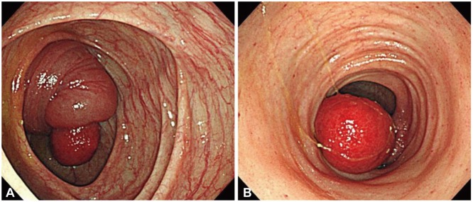
Fig. 1 (A) Colonoscopy showing an ileal lipoma with ileocolic intussusceptions through the ileocecal valve. (B) After air inflation, the ileal lipoma has reverted back to its original site in the ileum and the ileal mucosae have multiple hemorrhagic spots due to having been intussuscepted.
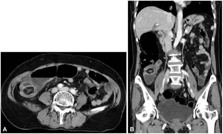
Fig. 2 (A) Abdominal computed tomography, showing a well-demarcated lipoma of 2.6×2.7 cm in size with a short peduncle in the terminal ileum. (B) Coronal section.
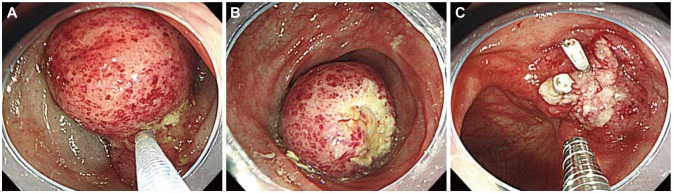
Fig. 3 (A) Submucosal injection of a mixture of saline and epinephrine into the peduncle of the ileal mass. (B) Snare polypectomy was performed by cap-assisted colonoscopy technique. (C) Hemoclipping at the base of the resected portion of the ileum to prevent delayed hemorrhage and perforation.
Reference
-
1. Begos DG, Sandor A, Modlin IM. The diagnosis and management of adult intussusception. Am J Surg. 1997; 173:88–94. PMID: 9074370.
Article2. Hancock BJ, Vajcner A. Lipomas of the colon: a clinicopathologic review. Can J Surg. 1988; 31:178–181. PMID: 3284624.3. Olmsted WW, Ros PR, Hjermstad BM, McCarthy MJ, Dachman AH. Tumors of the small intestine with little or no malignant predisposition: a review of the literature and report of 56 cases. Gastrointest Radiol. 1987; 12:231–239. PMID: 3596141.
Article4. Mayo CW, Pagtalunan RJ, Brown DJ. Lipoma of the alimentary tract. Surgery. 1963; 53:598–603. PMID: 13934160.5. Marinis A, Yiallourou A, Samanides L, et al. Intussusception of the bowel in adults: a review. World J Gastroenterol. 2009; 15:407–411. PMID: 19152443.
Article6. Thompson WM. Imaging and findings of lipomas of the gastrointestinal tract. AJR Am J Roentgenol. 2005; 184:1163–1171. PMID: 15788588.
Article7. Yoshimura H, Murata K, Takase K, Nakano T, Tameda Y. A case of lipoma of the terminal ileum treated by endoscopic removal. Gastrointest Endosc. 1997; 46:461–463. PMID: 9402125.
Article8. Son HS, Cho YS, Kim JS, et al. Endoscopic resection of a large colonic lipoma. Korean J Gastrointest Endosc. 2008; 37:122–126.9. Shin YK, Kim EY, Jeon SW, et al. A case of giant colonic lipoma endoscopically removed using an unroofing technique in phases. Korean J Gastrointest Endosc. 2008; 36:242–247.
- Full Text Links
-
- Actions
-
Cited
- CITED
-
- Close
- Share
-
- Similar articles
-
- Laparoscopic-assisted resection of ileal lipoma causing ileo-ileo-colic intussusception
- Efficacy of Total Colonoscopy with a Transparent Cap
- A Case of Giant Colonic Lipoma Showing Spontaneous Resolution after Endoscopic Partial Resection
- Painless Colonoscopy: Available Techniques and Instruments
- Comparison of the Complications and Urodynamic Parameters for Orthotopic Bladder Substitution with using Ileocolic or Ileal Segments after Radical Cystectomy
