Ultrasonographic Interval Changes in Solid Thyroid Nodules after Ultrasonography-Guided Fine-Needle Aspiration
- Affiliations
-
- 1Department of Radiology, Busan Paik Hospital, Inje University College of Medicine, Busan 47392, Korea. dwultra@nate.com
- 2Department of Pathology, Busan Paik Hospital, Inje University College of Medicine, Busan 47392, Korea.
- 3Department of Radiology, Gyeongsang National University Changwon Hospital, Gyeongsang National University School of Medicine, Changwon 51472, Korea.
- KMID: 2425122
- DOI: http://doi.org/10.3348/kjr.2018.19.1.158
Abstract
OBJECTIVE
None of the previous studies have investigated the interval change in ultrasonography (US) features of solid thyroid nodules (STNs) after US-guided fine-needle aspiration (US-FNA). This study aimed to assess the prevalence and characteristics of US interval changes in STNs after US-FNA.
MATERIALS AND METHODS
This study included 257 STNs in 257 patients in whom thyroid US and initial US-FNA had been performed by two radiologists from January 2015 to June 2015. One of the radiologists performed single needle puncture in all cases, whereas the other radiologist used double or triple needle punctures. Follow-up US examinations were performed after 12.0 ± 6.0 months. We evaluated the prevalence and characteristics of post-FNA US interval changes through a retrospective analysis. In addition, multiple factors were correlated with post-FNA US interval changes.
RESULTS
The number of needle punctures was one (n = 91), two (n = 163), and three (n = 3). Of the 257 STNs (mean diameter, 11.9 mm) in 257 patients, 35 (13.6%) showed an interval change in US features on follow-up US. Among them, 17 STNs (6.6%) showed newly developed malignant US features, including hypoechogenicity (n = 5), microcalcifications (n = 2), a spiculated margin (n = 4), hypoechogenicity with a spiculated margin (n = 5), and microcalcifications with non-parallel orientation (n = 1). Between patients who showed presence and absence of US interval changes, there were no significant differences in patient age, sex, nodule size, dichotomization, and location, Korean Thyroid Imaging Reporting and Data System categorization after FNA, practitioners involved, number of needle punctures, cytological findings, and interval between FNA and US follow-up (p > 0.05).
CONCLUSION
Awareness of US interval changes after US-FNA of STNs may be helpful for the management of STNs.
Keyword
MeSH Terms
Figure
-
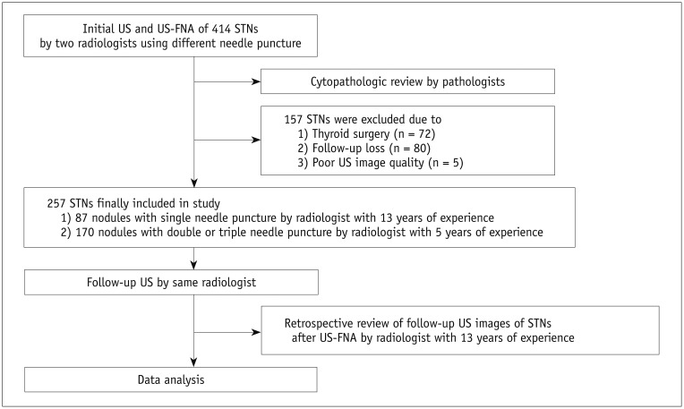
Fig. 1 Diagram illustrating enrollment of subjects and data analysis using study's algorithm.STNs = solid thyroid nodules, US = ultrasonography, US-FNA = US-guided fine-needle aspiration
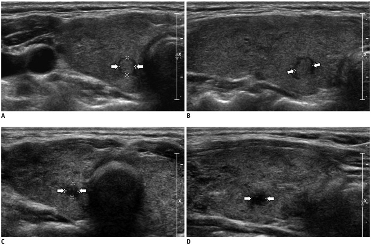
Fig. 2 55-year-old woman showing interval changes in US features of STN after US-FNA performed using double needle puncture.Before US-FNA, transverse (A) and longitudinal (B) gray-scale sonograms show STN with round shape in right thyroid lobe (arrows, 4.5 × 4.7 × 5.4 mm; K-TIRADS category 3). Radiologist with 5 years of experience performed US-FNA of this nodule, using double needle puncture. Cytological examination revealed that nodule belonged to Bethesda class II. At 12-month follow-up US after US-FNA, transverse (C) and longitudinal (D) gray-scale sonograms showed US interval changes in nodule (arrows). This nodule showed hypoechogenicity and spiculated margin, whereas nodule size decreased (3.7 × 3.8 × 4.6 mm; K-TIRADS category 5). Cytological examination after repeated US-FNA of this nodule revealed Bethesda class II. K-TIRADS = Korean Thyroid Imaging Reporting and Data System

Fig. 3 56-year-old woman with interval changes in US features of STN after US-FNA performed using single needle puncture.Before US-FNA, transverse (A) and longitudinal (B) gray-scale sonograms show STN with benign US features in left thyroid lobe (arrows, 6.0 × 10.4 × 11.2 mm; K-TIRADS category 3). Radiologist with 13 years of experience performed US-FNA of this nodule using single needle puncture. Cytological examination revealed that nodule belonged to Bethesda class II. At 12-month follow-up US after US-FNA, transverse (C) and longitudinal (D) gray-scale sonograms show US interval changes in nodule (arrows). This nodule showed decreased echogenicity and lobulated margin, whereas nodule size decreased (5.9 × 8.4 × 9.6 mm; K-TIRADS category 5). On same day, repeated US-FNA was performed for this nodule. Cytological examination revealed that nodule belonged to Bethesda class II.
Reference
-
1. Frates MC, Benson CB, Charboneau JW, Cibas ES, Clark OH, Coleman BG, et al. Management of thyroid nodules detected at US: Society of Radiologists in Ultrasound consensus conference statement. Radiology. 2005; 237:794–800. PMID: 16304103.
Article2. Shin JH, Baek JH, Chung J, Ha EJ, Kim JH, Lee YH, et al. Ultrasonography diagnosis and imaging-based management of thyroid nodules: Revised Korean Society of Thyroid Radiology consensus statement and recommendations. Korean J Radiol. 2016; 17:370–395. PMID: 27134526.
Article3. Ha EJ, Moon WJ, Na DG, Lee YH, Choi N, Kim SJ, et al. A Multicenter prospective validation study for the Korean thyroid imaging reporting and data system in patients with thyroid nodules. Korean J Radiol. 2016; 17:811–821. PMID: 27587972.
Article4. Haugen BR, Alexander EK, Bible KC, Doherty GM, Mandel SJ, Nikiforov YE, et al. 2015 American Thyroid Association Management Guidelines for adult patients with thyroid nodules and differentiated thyroid cancer: The American Thyroid Association Guidelines Task Force on thyroid nodules and differentiated thyroid cancer. Thyroid. 2016; 26:1–133. PMID: 26462967.
Article5. Lee YH, Baek JH, Jung SL, Kwak JY, Kim JH, Shin JH, et al. Ultrasound-guided fine needle aspiration of thyroid nodules: a consensus statement by the Korean Society of Thyroid Radiology. Korean J Radiol. 2015; 16:391–401. PMID: 25741201.
Article6. Kim DW, Lee EJ, Kim SH, Kim TH, Lee SH, Kim DH, et al. Ultrasound-guided fine-needle aspiration biopsy of thyroid nodules: comparison in efficacy according to nodule size. Thyroid. 2009; 19:27–31. PMID: 19021460.
Article7. Baloch ZW, LiVolsi VA. Post fine-needle aspiration histologic alterations of thyroid revisited. Am J Clin Pathol. 1999; 112:311–316. PMID: 10478135.
Article8. LiVolsi VA, Merino MJ. Worrisome histologic alterations following fine-needle aspiration of the thyroid (WHAFFT). Pathol Annu. 1994; 29:99–120. PMID: 7936753.9. Kini SR. Post-fine-needle biopsy infarction of thyroid neoplasms: a review of 28 cases. Diagn Cytopathol. 1996; 15:211–220. PMID: 8955603.
Article10. Ko MS, Jeong KS, Shong YK, Gong GY, Baek JH, Lee JH. Collapsing benign cystic nodules of the thyroid gland: sonographic differentiation from papillary thyroid carcinoma. AJNR Am J Neuroradiol. 2012; 33:124–127. PMID: 22158923.
Article11. Kim DW. Benign lesions that mimic thyroid malignancy on ultrasound. Can Assoc Radiol J. 2015; 66:79–85. PMID: 24931046.
Article12. Lacout A, Chevenet C, Marcy PY. Mummified thyroid syndrome. AJR Am J Roentgenol. 2016; 206:837–845. PMID: 27003052.
Article13. Sohn YM, Kim EK, Moon HJ, Kim SJ, Kwak JY. Suspiciously malignant findings on ultrasound after fine needle aspiration biopsy in a thyroid nodule with initially benign ultrasound and cytologic result: to repeat or to follow-up. Clin Imaging. 2011; 35:470–475. PMID: 22040793.
Article14. Russ G. Risk stratification of thyroid nodules on ultrasonography with the French TI-RADS: description and reflections. Ultrasonography. 2016; 35:25–38. PMID: 26324117.
Article15. Lee HY, Baek JH, Ha EJ, Park JW, Lee JH, Song DE, et al. Malignant-looking thyroid nodules with size reduction: core needle biopsy results. Ultrasonography. 2016; 35:327–334. PMID: 27184652.
Article16. Kim DW, Choo HJ, Park JS, Lee EJ, Kim SH, Jung SJ, et al. Ultrasonography-guided fine-needle aspiration cytology for thyroid nodules: an emphasis on one-sampling and biopsy techniques. Diagn Cytopathol. 2012; 40(Suppl 1):E48–E54. PMID: 21416648.
Article17. Cibas ES, Ali SZ. The Bethesda system for reporting thyroid cytopathology. Thyroid. 2009; 19:1159–1165. PMID: 19888858.
Article
- Full Text Links
-
- Actions
-
Cited
- CITED
-
- Close
- Share
-
- Similar articles
-
- Thyroid nodules with discordant results of ultrasonographic and fine-needle aspiration findings
- Indications for Fine Needle Aspiration in Thyroid Nodules
- Natural Course of Cytologically Diagnosed Benign Thyroid Nodules
- An Analysis of the Ultrasound Findings of False Negative Cases for an Initial Ultrasound-guided Fine Needle Aspiration Biopsy (FNAB)
- Follow-up of benign thyroid nodules confirmed by ultrasound-guided core needle biopsy after inconclusive cytology on fine-needle aspiration biopsy
