Size-Specific Dose Estimation In the Korean Lung Cancer Screening Project: Does a 32-cm Diameter Phantom Represent a Standard-Sized Patient in Korean Population?
- Affiliations
-
- 1Department of Radiology, Gachon University Gil Medical Center, Incheon 21565, Korea.
- 2Department of Radiology and Center for Imaging Science, Samsung Medical Center, Sungkyunkwan University School of Medicine, Seoul 06351, Korea. taejung.kim1@gmail.com
- 3Department of Radiology, Seoul National University College of Medicine, Seoul 03080, Korea.
- 4Department of Diagnostic Radiology, National Cancer Center, Goyang 10408, Korea.
- 5Department of Radiology, Pusan National University School of Medicine and Medical Research Institute, Pusan National University Hospital, Busan 49241, Korea.
- 6Department of Radiology, Chungbuk National University Hospital, Cheongju 28644, Korea.
- 7Cancer Early Detection Branch, National Cancer Control Institute, National Cancer Center, Goyang 10408, Korea.
- KMID: 2424857
- DOI: http://doi.org/10.3348/kjr.2018.19.6.1179
Abstract
OBJECTIVE
The purposes of this study were to evaluate size-specific dose estimate (SSDE) of low-dose CT (LDCT) in the Korean Lung Cancer Screening (K-LUCAS) project and to determine whether CT protocols from Western countries are appropriate for lung cancer screening in Korea.
MATERIALS AND METHODS
For participants (n = 256, four institutions) of K-LUCAS pilot study, volume CT dose index (CTDI(vol)) using a 32-cm diameter reference phantom was compared with SSDE, which was recalculated from CTDI(vol) using size-dependent conversion factor (f-size) based on the body size, as described in the American Association of Physicists in Medicine Report 204. This comparison was subsequently assessed by body mass index (BMI) levels (underweight/normal vs. overweight/obese), and automatic exposure control (AEC) adaptation (yes/no).
RESULTS
Size-specific dose estimate was higher than CTDI(vol) (2.22 ± 0.75 mGy vs. 1.67 ± 0.60 mGy, p < 0.001), since the f-size was larger than 1.0 for all participants. The ratio of SSDE to CTDI(vol) was higher in lower BMI groups; 1.26, 1.37, 1.43, and 1.53 in the obese (n = 103), overweight (n = 70), normal (n = 75), and underweight (n = 4), respectively. The ratio of SSDE to CTDI(vol) was greater in standard-sized participants than in large-sized participants independent of AEC adaptation; with AEC, SSDE/CTDI(vol) in large- vs. standard-sized participants: 1.30 ± 0.08 vs. 1.44 ± 0.08 (p < 0.001) and without AEC, 1.32 ± 0.08 vs. 1.42 ± 0.06 (p < 0.001).
CONCLUSION
Volume CT dose index based on a reference phantom underestimates radiation exposure of LDCT in standard-sized Korean participants. The optimal radiation dose limit needs to be verified for standard-sized Korean participants.
Keyword
MeSH Terms
Figure
-
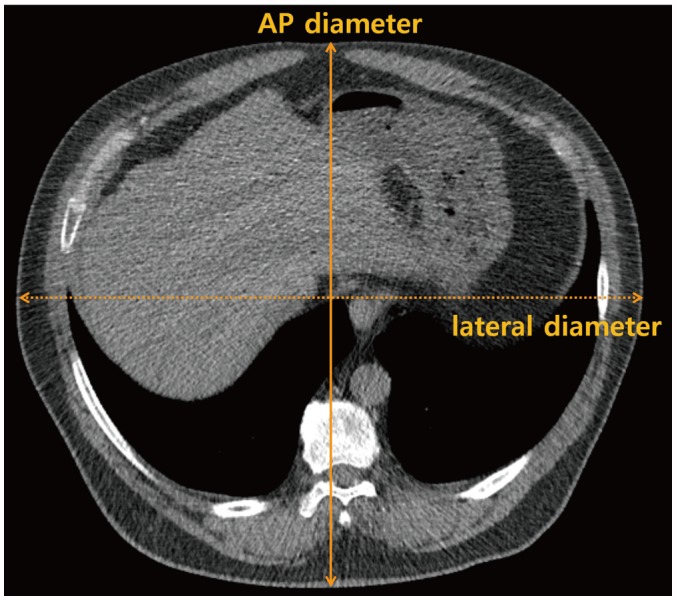
Fig. 1 Body size measurement using low-dose screening chest CT. AP diameter and lateral diameter are measured at mid-liver level.AP = antero-posterior
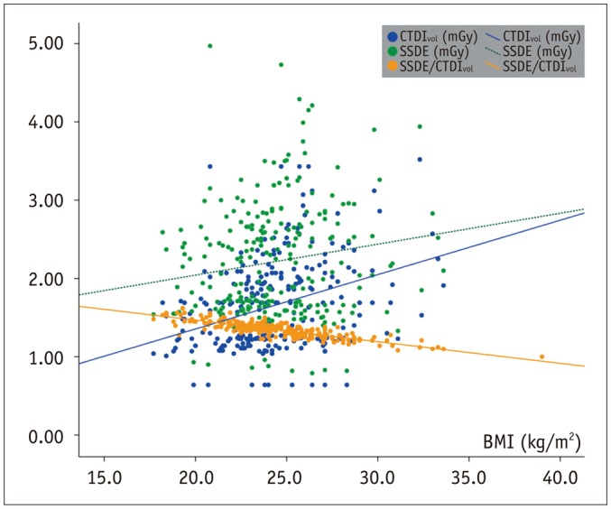
Fig. 2 Distribution of radiation dose estimates based on 32-cm diameter reference phantom (CTDIvol), SSDEs, and SSDE/CTDIvol according to BMI.BMI = body mass index, CTDIvol = volume CT dose index, SSDE = size-specific dose estimate, SSDE/CTDIvol = ratio of SSDE to CTDIvol
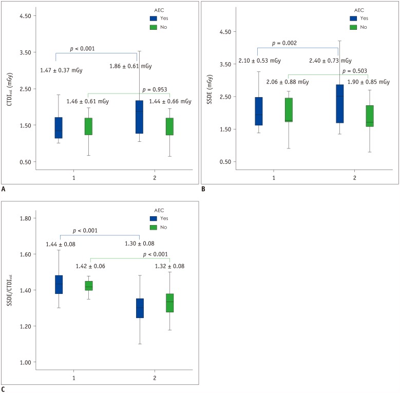
Fig. 3 Comparison of radiation dose estimates according to BMI and use of AEC.A. Radiation dose estimate based on 32-cm diameter reference phantom (CTDIvol). B. SSDEs. C. SSDE/CTDIvol according to BMI and AEC. AEC = automatic exposure control, 1 = underweight and normal, 2 = overweight and obese
Cited by 1 articles
-
Rise of the Visible Monkey: Sectioned Images of Rhesus Monkey
Beom Sun Chung, Chang-Yeop Jeon, Jae-Won Huh, Kang-Jin Jeong, Donghwan Har, Kyu-Sung Kwack, Jin Seo Park
J Korean Med Sci. 2019;34(8):. doi: 10.3346/jkms.2019.34.e66.
Reference
-
1. Kramer BS, Berg CD, Aberle DR, Prorok PC. Lung cancer screening with low-dose helical CT: results from the National Lung Screening Trial (NLST). J Med Screen. 2011; 18:109–111. PMID: 22045816.
Article2. Lee JW, Kim HY, Goo JM, Kim EY, Lee SJ, Kim TJ, et al. Radiological report of pilot study for the Korean Lung Cancer Screening (K-LUCAS) project: feasibility of implementing lung imaging reporting and data system. Korean J Radiol. 2018; 19:803–808. PMID: 29962887.
Article3. McNitt-Gray MF. AAPM/RSNA physics tutorial for residents: topics in CT. Radiation dose in CT. Radiographics. 2002; 22:1541–1553. PMID: 12432127.4. WHO Expert Consultation. Appropriate body-mass index for Asian populations and its implications for policy and intervention strategies. Lancet. 2004; 363:157–163. PMID: 14726171.5. Larke FJ, Kruger RL, Cagnon CH, Flynn MJ, McNitt-Gray MM, Wu X, et al. Estimated radiation dose associated with low-dose chest CT of average-size participants in the National Lung Screening Trial. AJR Am J Roentgenol. 2011; 197:1165–1169. PMID: 22021510.
Article6. Aberle DR, Gamsu G, Henschke CI, Naidich DP, Swensen SJ. A consensus statement of the Society of Thoracic Radiology: screening for lung cancer with helical computed tomography. J Thorac Imaging. 2001; 16:65–68. PMID: 11149694.7. AAPM Task Group 23 of the Diagnostic Imaging Council CT Committee. The measurement, reporting and management of radiation dose in CT (Report No. 96). College Park, MD: American Association of Physicists in Medicine;2008.8. AAPM Task Group 204. Size-specific dose estimates (SSDE) in pediatric and adult body CT examinations. College Park, MD: American Association of Physicists in Medicine;2008.9. McCollough CH, Leng S, Yu L, Cody DD, Boone JM, McNitt-Gray MF. CT dose index and patient dose: they are not the same thing. Radiology. 2011; 259:311–316. PMID: 21502387.10. Christner JA, Braun NN, Jacobsen MC, Carter RE, Kofler JM, McCollough CH. Size-specific dose estimates for adult patients at CT of the torso. Radiology. 2012; 265:841–847. PMID: 23091173.
Article
