Risk Factors for Hinge Fracture Associated with Surgery Following Cervical Open-Door Laminoplasty
- Affiliations
-
- 1Department of Neurosurgery, Pusan National University Hospital, Pusan National University School of Medicine, Busan, Korea.
- 2Catholic Neuroscience Center, Department of Neurosurgery, Yeouido St. Mary's Hospital, The Catholic University College of Medicine, Seoul, Korea. chough@catholic.ac.kr
- KMID: 2424323
- DOI: http://doi.org/10.13004/kjnt.2018.14.2.118
Abstract
OBJECTIVE
The purpose of this study was to analyze the risk factors for hinge fracture (HF) and non-union during cervical open-door laminoplasty (CODL).
METHODS
We included 25 patients who underwent CODL with available serial computed tomography scans acquired at 2 days and 1 year postoperatively. Patients' medical records and radiographic data were reviewed. Risk factors related to the surgical procedures were evaluated including the lamina angle, spinous angle, difference in the lamina angle, outer cortex location (OCL), and inner cortex location.
RESULTS
There were a total of 76 hinges. Of these, 44 laminae were classified as deformed hinges, and 32 were classified as fragmented hinges. Additionally, 66 laminae were healed completely, and 10 laminae were not healed by 12 months postoperatively. The OCL (odds ratio, 70.45; 95% confidence interval, 7.73-641.76) was identified as a predictor of HFs immediately following CODL. However, none of the factors we evaluated was related to hinge non-union.
CONCLUSION
A medially located hinge gutter ( >1.9 mm from the pedicle on the outer cortex) seems to be an important risk factor for HFs following CODL. However, the hinge healing status was not related to the surgical technique.
Figure
-

FIGURE 1 (A) Schematic diagram of the lamina angle (LA) and spinous angle (SA). SA (a) was defined as the angle between the parallel line to spinous process and the perpendicular line to virtual line crossing both lateral mass posteriorly. LA (b) was defined as the angle between parallel line to the hinge side lamina and virtual line crossing both lateral mass posteriorly. (B) Radiographic measurements of hinge gutter location. Inner cortex layer (c) was defined as distance from pedicle to inner cortical bone of hinge, and outer cortex layer (d) was defined as distance from pedicle to outer cortical bone of hinge.
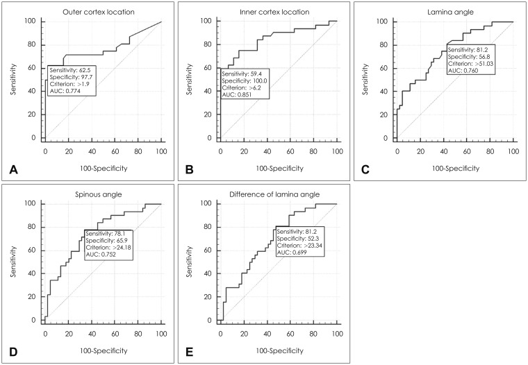
FIGURE 2 Receiver-operating characteristics curves of risk factors. (A) Outer cortex location, (B) inner cortex location, (C) lamina angle, (D) spinous angle, and (E) difference of lamina angle. AUC: area under curve.
Reference
-
1. Byard RW, Langlois N, Gilbert JD. Positive “water test”-an external indicator of base of skull hinge-ring fracture. J Forensic Sci. 2010; 55:519–520. PMID: 20102468.
Article2. Chen H, Liu H, Zou L, Li T, Gong Q, Song Y, et al. Effect of miniplate fixation on hinge fracture and bony fusion in unilateral open-door cervical expansive laminoplasty. Clin Spine Surg. 2016; 29:E288–E295. PMID: 25023712.
Article3. Cho SH, Lee JH, Chough CK, Joo WI, Park HK, Lee KJ, et al. Hinge Fracture during Cervical Open-door Laminoplasty: Does it Affect Clinical and Radiographic Outcomes? Korean J Spine. 2014; 11:45–51. PMID: 25110482.
Article4. Hirabayashi K, Satomi K. Operative procedure and results of expansive open-door laminoplasty. Spine (Phila Pa 1976). 1988; 13:870–876. PMID: 3143157.
Article5. Hur JW, Park YK, Kim BJ, Moon HJ, Kim JH. Risk factors for delayed hinge fracture after plate-augmented cervical open-door laminoplasty. J Korean Neurosurg Soc. 2016; 59:368–373. PMID: 27446518.
Article6. Lee S, Chung CK, Kim CH. Risk factor analysis of hinge fusion failure after plate-only open-door laminoplasty. Global Spine J. 2015; 5:9–16. PMID: 25648062.
Article7. O'Brien MF, Peterson D, Casey AT, Crockard HA. A novel technique for laminoplasty augmentation of spinal canal area using titanium miniplate stabilization. A computerized morphometric analysis. Spine (Phila Pa 1976). 1996; 21:474–483. PMID: 8658252.
- Full Text Links
-
- Actions
-
Cited
- CITED
-
- Close
- Share
-
- Similar articles
-
- Risk Factors for Delayed Hinge Fracture after Plate-Augmented Cervical Open-Door Laminoplasty
- Hinge Fracture during Cervical Open-door Laminoplasty: Does it Affect Clinical and Radiographic Outcomes?
- Comparison of Midline Splitting versus Unilateral Open Door Laminoplasty and Its Impact on Patient Outcomes
- Comparison of Clinical Results according to the Complications after or during Open Door Laminoplasty Surgery for Cervical Myelopathy
- Cervical Open-Door Laminoplasty by Hydroxyapatite Implant Insertion Without Suturing
