Delayed Endovascular Coil Extrusion Presenting as a Foreign Body of the Throat: a Case Report
- Affiliations
-
- 1Department of Otorhinolaryngology and Head and Neck Surgery, All India Institute of Medical Sciences, New Delhi, India. kapil_sikka@yahoo.com
- 2Department of Neuroimaging and Interventional Neuroradiology, All India Institute of Medical Sciences, New Delhi, India.
- KMID: 2424053
- DOI: http://doi.org/10.5469/neuroint.2018.13.1.66
Abstract
- Endovascular treatment is a standard mode of treatment for traumatic cavernous internal carotid artery (ICA) pseudoaneurysms with good results and relatively low rates of complications. We describe a case of an unusual, potentially fatal, delayed postoperative event happening in a case of post-traumatic pseudoaneurysm of ICA, which had been previously managed with endovascular coiling.
Keyword
Figure
-

Fig. 1 Left ICA angiogram (A) reveals traumatic left cavernous ICA pseudoaneurysm, which was subsequently treated by coil embolization (B). Final left CCA angiogram (C) shows complete occlusion of both aneurysm and parent artery. Follow up angiogram (D & E) 1 year later shows stable occlusion of both aneurysm and parent artery, however, loosening of coil mass along its anterior aspect is noted (arrow in E). Two years after embolization, left ICA angiogram (F) shows extrusion of coil loop into oropharynx (star) with stable aneurysm occlusion.

Fig. 2 Lateral skull radiograph (A) immediately after embolization reveals compact coil mass in region of pseudoaneurysm. Follow up radiograph (B) after 1 year shows loosening of coil mass along its anterior aspect (red arrow in B). Lateral radiograph (C) and reformatted sagittal CT (D) on two years after embolization show extrusion of coil loop into oropharynx (blue arrows in C & D). Final radiograph (E) and reformatted sagittal CT (F), after post endoscopic excision of the herniated coil loop.
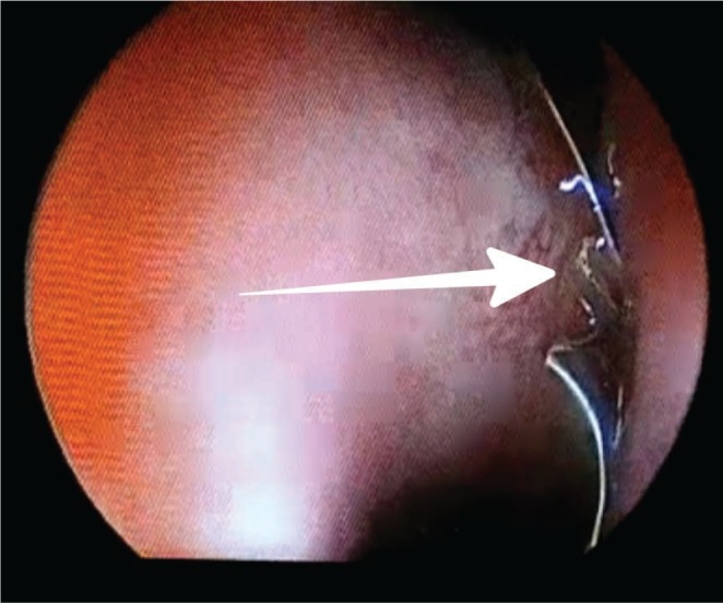
Fig. 3 Closer view inside nasal cavity showing the part of the coil (white arrow) behind middle turbinate.
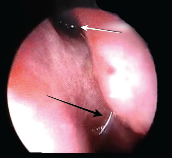
Fig. 4 Nasal endoscopy showing the coil in the nasopharynx (Black arrow pointing to the part in choana and the white arrow showing the part in sphenoethmoid recess).

Fig. 5 Coil wire being retrieved endoscopically.
Reference
-
1. Chen D, Concus AP, Halbach VV, Cheung SW. Epistaxis originating from traumatic pseudoaneurysm of the internal carotid artery: diagnosis and endovascular therapy. Laryngoscope. 1998; 108:326–331. PMID: 9504602.
Article2. Higashida RT, Halbach VV, Dowd CF, Barnwell SL, Hieshima GB. Intracranial aneurysms: interventional neurovascular treatment with detachable balloons-results in 215 cases. Radiology. 1991; 178:663–670. PMID: 1994399.
Article3. Maras D, Lioupis C, Magoufis G, Tsamopoulos N, Moulakakis K, Andrikopoulos V. Covered stent-graft treatment of traumatic internal carotid artery pseudoaneurysms: a review. Cardiovasc Intervent Radiol. 2006; 29:958–968. PMID: 16897263.
Article4. Levy E, Koebbe CJ, Horowitz MB, Pride GL, Dutton K, et al. Rupture of intracranial aneurysms during endovascular coiling: management and outcomes. Neurosurgery. 2001; 49:807–811. PMID: 11564240.
Article5. Akan H, Belet U, Enel A. Coil-Induced Perforation of Recently Ruptured Cerebral Aneurysm during Embolization. Causes and Avoidance. Interv Neuroradiol. 2003; 9:83–88.
- Full Text Links
-
- Actions
-
Cited
- CITED
-
- Close
- Share
-
- Similar articles
-
- Delayed cranial nerve palsy after successful coil embolization in cavernous sinus lesion
- Oral Extrusion of Screw after Anterior Cervical Interbody Fusion
- Extrusion of Gutta-Percha into the Nasal Cavity Causing Maxillary Fungal Sinusitis: A Case Report
- Acute frame coil migration during filling coil retrieval in a cerebral aneurysm embolization case: A possible result of a venturi effect?
- Delayed Herniation of Coil Loop and Spontaneous Reposition in a Superior Cerebellar Artery Aneurysm
