Secondary Aneurysmal Bone Cyst in a Craniofacial Fibrous Dysplasia: Case Report
- Affiliations
-
- 1Department of Neurosurgery, Konkuk University Medical Center, Seoul, Korea. kohyc@kuh.ac.kr
- 2Department of Radiology, Konkuk University Medical Center, Seoul, Korea.
- 3Department of Pathology, Konkuk University Medical Center, Seoul, Korea.
- KMID: 2423980
- DOI: http://doi.org/10.14791/btrt.2018.6.e15
Abstract
- Aneurysmal bone cyst (ABC) is a rare non-neoplastic bone lesion that involves mostly the long bones and vertebrae and may occur very rarely in the craniofacial bones. ABCs may occur as secondary bony pathologies in association with various benign and malignant bone tumors and with fibrous dysplasia (FD). FD is a common non-neoplastic bony pathology mostly affecting craniofacial bones. Secondary ABC occurring in craniofacial FD is extremely rare, with only approximately 20 cases reported in the literature to date. Here, we report on a case of secondary ABC in a 25-year-old woman who has had a craniofacial deformity for over 10 years and who presented to us with a rapidly growing painful pulsatile mass in the right frontal region that began over 2 months prior to admission. On thorough examination of computed tomography and magnetic resonance imaging brain scans taken at two-month interval, an aggressive, rapidly enlarging ABC, arising from the right frontal FD, was diagnosed. The patient underwent preoperative embolization followed by gross total resection of the ABC and cranioplasty. The 6-month follow up showed no recurrence of the ABC, nor was any progression of the FD noticed.
MeSH Terms
Figure
-

Fig. 1 Preoperative CT scan of the patient; axial (A), coronal (B), and sagittal (C) image. Image shows diffuse fibrous dysplasia in the frontal bone with an osteolytic lesion.

Fig. 2 MRI, gadolinium-enhanced T1-weighted image. Extensive fibrous dysplasia involving frontal, sphenoid, and ethmoid bones, midline parietal bones, both temporal bones, midline and left occipital bone, clivus, and right zygomatic bone (A: axial image, B: coronal image, C: sagittal image). Fluid-fluid level with a large aneurysmal bone cyst (6.2×5.5×5.0 cm) was seen in the right frontal bone (D: axial image, E: coronal image, F: sagittal image).
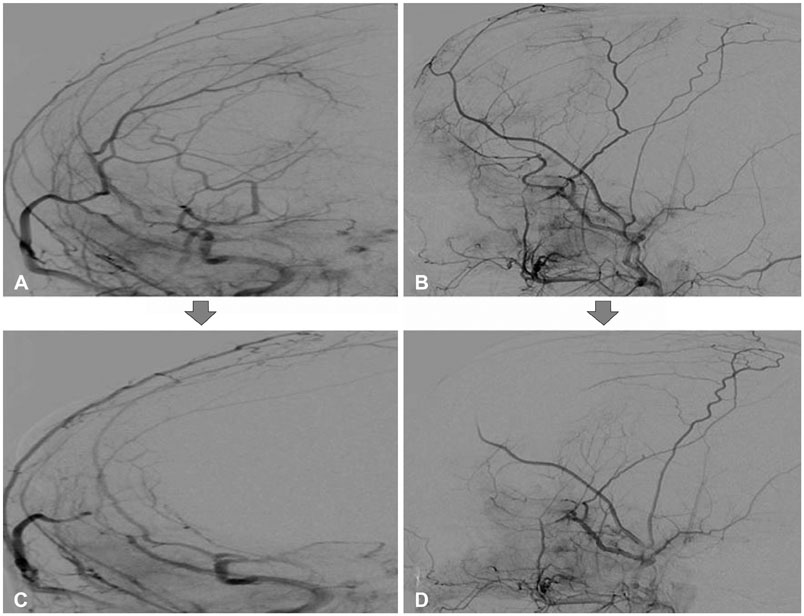
Fig. 3 Trans-femoral cerebral angiography image of the patient. The pre-embolization image showed prominent vascular stain in the right frontal area, at the margin of the aneurysmal bone cyst (A: anterior-posterior view, B: right lateral view). An endovascular embolization was performed in a branch of the right middle meningeal artery and the right superficial temporal artery. After embolization, vascular staining was markedly decreased, but residual staining was seen in the posterior and inferior margins (C: anterior-posterior view, D: right lateral view).

Fig. 4 Histological examination of the resected lesion. A: The aneurysmal bone cyst wall (hematoxylin and eosin staining, original magnification ×100). B: A multinucleated osteoclastic giant cell (hematoxylin and eosin staining, original magnification ×200). C: Woven bone with proliferating fibrous tissue (hematoxylin and eosin staining, original magnification ×200).

Fig. 5 Postoperative axial CT scan of the setting bone shows the partial removal of the fibrous dysplasia around the aneurysmal bone cysts (A and B).

Fig. 6 Cosmetic improvement of the patient's forehead (A: preoperative image, B: postoperative image).
Reference
-
1. Edgerton MT, Persing JA, Jane JA. The surgical treatment of fibrous dysplasia. With emphasis on recent contributions from cranio-maxillo-facial surgery. Ann Surg. 1985; 202:459–479.2. Casselman JW, De Jonge I, Neyt L, De Clercq C, D'Hont G. MRI in craniofacial fibrous dysplasia. Neuroradiology. 1993; 35:234–237.
Article3. Mattei TA, Mattei JA, Ramina R, Aguiar PH. Fibrous dysplasia in combination with aneurysmal bone cyst presenting as a subarachnoid haemorrhage. Neurol Sci. 2005; 26:178–181.
Article4. Dumont AS, Boulos PT, Jane JA Jr, Ellegala DB, Newman SA, Jane JA Sr. Cranioorbital fibrous dysplasia: with emphasis on visual impairment and current surgical management. Neurosurg Focus. 2001; 10:E6.
Article5. Maher CO, Friedman JA, Meyer FB, Lynch JJ, Unni K, Raffel C. Surgical treatment of fibrous dysplasia of the skull in children. Pediatr Neurosurg. 2002; 37:87–92.
Article6. Chong VF, Khoo JB, Fan YF. Fibrous dysplasia involving the base of the skull. AJR Am J Roentgenol. 2002; 178:717–720.
Article7. Ruggieri P, Sim FH, Bond JR, Unni KK. Malignancies in fibrous dysplasia. Cancer. 1994; 73:1411–1424.
Article8. Tuna H, Karatas A, Yilmaz ER, Yagmurlu B, Erekul S. Aneurysmal bone cyst of the temporal bone: case report. Surg Neurol. 2003; 60:571–574.
Article9. Lin WC, Wu HT, Wei CJ, Chang CY. Aneurysmal bone cyst arising from fibrous dysplasia of the frontal bone (2004:2b). Eur Radiol. 2004; 14:930–932.
Article10. Branch CL Jr, Challa VR, Kelly DL Jr. Aneurysmal bone cyst with fibrous dysplasia of the parietal bone. Report of two cases. J Neurosurg. 1986; 64:331–335.11. Rappaport ZH. Aneurysmal bone cyst associated with fibrous dysplasia of the skull. Neurochirurgia (Stuttg). 1989; 32:192–194.
Article12. Wojno KJ, McCarthy EF. Fibro-osseous lesions of the face and skull with aneurysmal bone cyst formation. Skeletal Radiol. 1994; 23:15–18.
Article13. Haddad GF, Hambali F, Mufarrij A, Nassar A, Haddad FS. Concomitant fibrous dysplasia and aneurysmal bone cyst of the skull base. Case report and review of the literature. Pediatr Neurosurg. 1998; 28:147–153.
Article14. Saito I, Asano T, Sano K, et al. Neuroprotective effect of an antioxidant, ebselen, in patients with delayed neurological deficits after aneurysmal subarachnoid hemorrhage. Neurosurgery. 1998; 42:269–277.
Article15. Itshayek E, Spector S, Gomori M, Segal R. Fibrous dysplasia in combination with aneurysmal bone cyst of the occipital bone and the clivus: case report and review of the literature. Neurosurgery. 2002; 51:815–817.
Article16. Pasquini E, Ceroni Compadretti G, Sciarretta V, Ippolito A. Transnasal endoscopic surgery for the treatment of fibrous dysplasia of maxillary sinus associated to aneurysmal bone cyst in a 5-year-old child. Int J Pediatr Otorhinolaryngol. 2002; 62:59–62.
Article17. Iseri PK, Efendi H, Demirci A, Komsuoglu S. Fibrous dysplasia of the cranial bones: a case report and review of the literature. Yale J Biol Med. 2005; 78:141–145.18. Lee JW, Kim JH, Han SH, Kang HI. Fibrous dysplasia with aneurysmal bone cyst presenting as painful solitary skull lesion. J Korean Neurosurg Soc. 2010; 48:551–554.
Article19. Terkawi AS, Al-Qahtani KH, Baksh E, Soualmi L, Mohamed Ael-B, Sabbagh AJ. Fibrous dysplasia and aneurysmal bone cyst of the skull base presenting with blindness: a report of a rare locally aggressive example. Head Neck Oncol. 2011; 3:15.
Article20. Manjila S, Zender CA, Weaver J, Rodgers M, Cohen AR. Aneurysmal bone cyst within fibrous dysplasia of the anterior skull base: continued intracranial extension after endoscopic resections requiring craniofacial approach with free tissue transfer reconstruction. Childs Nerv Syst. 2013; 29:1183–1192.
Article21. Hnenny L, Roundy N, Zherebitskiy V, Grafe M, Mansoor A, Dogan A. Giant aneurysmal bone cyst of the anterior cranial fossa and paranasal sinuses presenting in pregnancy: case report and literature review. J Neurol Surg Rep. 2015; 76:e216–e221.
Article22. Davies AM, Evans N, Mangham DC, Grimer RJ. MR imaging of brown tumour with fluid-fluid levels: a report of three cases. Eur Radiol. 2001; 11:1445–1449.
Article23. Nguyen BD, Lugo-Olivieri CH, McCarthy EF, Frassica FJ, Ma LD, Zerhouni EA. Fibrous dysplasia with secondary aneurysmal bone cyst. Skeletal Radiol. 1996; 25:88–91.
Article24. Cakirer S, Basak M, Celebi I, Kabukcuoglu F, Erdem Y. Aneurysmal bone cyst of the temporal bone. Curr Probl Diagn Radiol. 2003; 32:169–175.
Article25. Chidambaram B, Santosh V, Balasubramaniam V. Aneurysmal bone cyst of the temporal bone. Childs Nerv Syst. 2001; 17:411–414.
Article26. Lippman CR, Jallo GI, Feghali JG, Jimenez E, Epstein F. Aneurysmal bone cyst of the temporal bone. Pediatr Neurosurg. 1999; 31:219–223.
Article27. O'Brien DP, Rashad EM, Toland JA, Farrell MA, Phillips J. Aneurysmal cyst of the frontal bone: case report and review of the literature. Br J Neurosurg. 1994; 8:105–108.
- Full Text Links
-
- Actions
-
Cited
- CITED
-
- Close
- Share
-
- Similar articles
-
- Fibrous Dysplasia with Aneurysmal Bone Cyst Presenting as Painful Solitary Skull lesion
- Aneurysmal bone cyst arising from the surgically removed craniofacial fibrous dysplasia in the long-term follow-up: a case report
- A Case of Fibrous Dysplasia of the Ethmoid Bone Forming Intracranial Cyst
- Solitary Bone Cyst of the Proximal Femur Mimicking Fibrous Dysplasia: A Case Report
- Current concepts of craniofacial fibrous dysplasia: pathophysiology and treatment
