Avulsion Fracture of the Posterior Cruciate Ligament from Femoral Insertion Occurred in a Patient with Residual Poliomyelitis: A Case Report
- Affiliations
-
- 1Department of Orthopaedic Surgery, CHA Bundang Medical Center, CHA University School of Medicine, Seongnam, Korea. wcosdoc@gmail.com
- KMID: 2422896
- DOI: http://doi.org/10.12671/jkfs.2018.31.4.149
Abstract
- Avulsion fracture of the posterior cruciate ligament from its femoral insertion is quite rare, particularly in adults, and the treatment guidelines have not been established. A 68-year-old female patient with residual poliomyelitis presented with an avulsion fracture of the femoral insertion of the posterior cruciate ligament after a falling accident and was treated with arthroscopic headless compression screw fixation and pull-out suture of the avulsed ligament. We report this case with a relevant discussion of this type of injury.
Keyword
MeSH Terms
Figure
-
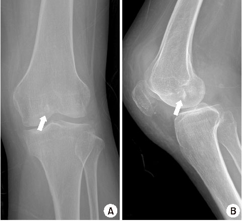
Fig. 1 Anteroposterior (A) and lateral (B) radiographs of the left knee showing a bony fragment (arrows) in the femur intercondylar notch area.

Fig. 2 Axial (A) and sagittal (B) computed tomography images of the left knee showing a comminuted fracture (white arrows) located at the inner aspect of the intercondylar notch at the lateral side of the medial femoral condyle. (C) Three-dimensional reconstruction of a computed tomography image shows a fracture located at the femoral insertion of the posterior cruciate ligament (black arrow).

Fig. 3 Sagittal fat-suppressed T2-weighted magnetic resonance image of the left knee showing an avulsion fracture of the posterior cruciate ligament (arrow). Increased bone marrow signal on inferior part of the patella (arrowhead) and increased joint effusion (asterisk) are also observed.

Fig. 4 Posterior drawer test of the left knee performed under anesthesia showing the starting point (A) and posterior displacement (B) of the tibia after a force was applied suggestive of posterior cruciate ligament insufficiency.
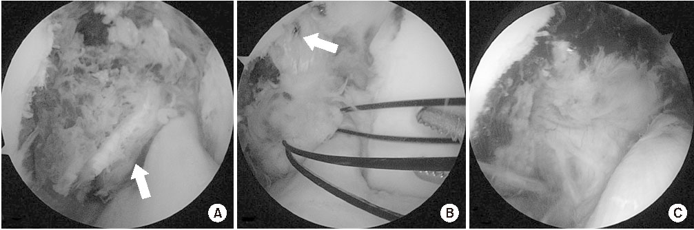
Fig. 5 Intraoperative arthroscopic findings of the left knee. (A) Avulsion fracture of the femur medial condyle was observed, and it was connected to the posterior cruciate ligament (arrow). (B) After fixation of the fragment using a headless compression screw (arrow), the avulsed posterior cruciate ligament is sutured for pull-out fixation. (C) Tension of the posterior cruciate ligament was confirmed using the arthroscopic probe after the fixation.
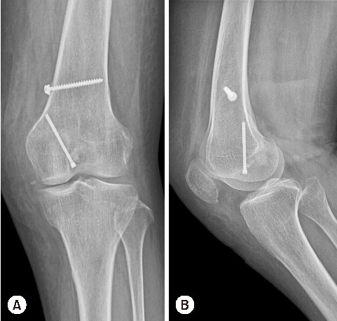
Fig. 6 Postoperative anteroposterior (A) and lateral (B) radiographs of the left knee. A headless compression screw was inserted in the medial femoral condyle and a cancellous screw with a washer was inserted into the metaphysis of the femur for post-tie fixation of the pull-out sutures.
Reference
-
1. Giordano BD, Dehaven KE, Maloney MD. Acute femoral “peel-off” tears of the posterior cruciate ligament: technique for arthroscopic anatomical repair. Am J Orthop (Belle Mead NJ). 2011; 40:226–232.2. Rosso F, Bisicchia S, Amendola A. Arthroscopic repair of “peel-off” lesion of the posterior cruciate ligament at the femoral condyle. Arthrosc Tech. 2014; 3:e149–e154.
Article3. Albtoush OM, Horger M. Insufficiency avulsion fracture of the femoral attachment of the posterior cruciate ligament of the knee joint. Skeletal Radiol. 2017; 46:1267–1269.
Article4. Lee YS, Ahn JH, Park JH, Park JW, Yoon JY, Kum DH. Partial femoral avulsion of the posterior cruciate ligament presenting as an osteochondral defect. J Knee Surg. 2009; 22:369–371.
Article5. Mishra AK, Vikas R. A rare case of bony avulsion of posterior cruciate ligament from its femoral attachment. Med J Armed Forces India. 2016; 72:S98–S100.
Article6. Park IS, Kim SJ. Arthroscopic fixation of avulsion of the posterior cruciate ligament from femoral insertion. Arthroscopy. 2005; 21:1397.
Article7. Xu Z, Chen D, Shi D, Jiang Q. Case report: osteochondral avulsion fracture of the posteromedial bundle of the PCL in knee hyperflexion. Clin Orthop Relat Res. 2012; 470:3616–3623.
Article8. Martini F, Kremling E, Sell S. Bilateral atraumatic avulsion fracture of the calcaneal tubercle in osteomalacia during fluoride therapy: a case report. Acta Orthop Scand. 1999; 70:91–92.
Article9. Onada Y, Umemoto T, Fukuda K, Kajino T. Coracoid process avulsion fracture at the coracoclavicular ligament attachment site in an osteoporotic patient with acromioclavicular joint dislocation. Case Rep Orthop. 2016; 2016:9580485.
Article10. Hegde V, Jo JE, Andreopoulou P, Lane JM. Effect of osteoporosis medications on fracture healing. Osteoporos Int. 2016; 27:861–871.
Article
- Full Text Links
-
- Actions
-
Cited
- CITED
-
- Close
- Share
-
- Similar articles
-
- Bilateral PCL Avulsion Fracture from Tibial Attatchment Site in a 16-years-old Male : A Case Report
- Avulsion of the Femoral Attachment of Anterior Cruciate Ligament Associated with Ipsilateral Femoral Shaft Fracture in Skeletally Mature Patient: A Case Report
- Avulsion Fractures of the Anterior Cruciate and Posterior Cruciate Ligaments in a Skeletally Immature Child
- Avulsion Fracture of Femoral Attachment of Anterior Cruciate Ligament in a 7-Year-Old Girl
- The Results of Primary Repair in Acute Injuries of the Posterior Cruciate Ligament
