Bilateral Infarction of the Recurrent Arteries of Heubner Following Clipping of an Anterior Communicating Artery Aneurysm
- Affiliations
-
- 1Department of Neurosurgery, Gyeongsang National University Hospital, Gyeongsang National University School of Medicine, Jinju, Korea. chl68@gnu.ac.kr
- KMID: 2422557
- DOI: http://doi.org/10.7461/jcen.2018.20.1.28
Abstract
- A 50-year-old woman reported to the emergency department with thunderclap headache and vomiting. Non-enhanced brain computed tomography (CT) showed a subarachnoid hemorrhage of Hunt-Hess Grade II and Fisher Grade III. Brain angiography CT and transfemoral cerebral angiography (TFCA) revealed an aneurysm of the anterior communicating artery. A direct neck clipping was performed using the pterional approach. The post-operation CT was uneventful. Six days postoperatively, the patient became lethargic. The mean velocity (cm/s) of the middle cerebral artery peaked at 173 cm/s on the right side and 167 cm/s on the left. A TFCA revealed decreased perfusion in both recurrent arteries of Heubner (RAH), but no occlusion in either. Intra-arterial nimodipine injection was administered. On the 7th postoperative day, CT demonstrated a newly developed low-density lesion in the RAH territory bilaterally. The cause of the infarction was attributed to decreased perfusion caused by cerebral vasospasm. The patient was discharged with no definite neurologic deficit except for mild cognitive disorder.
MeSH Terms
-
Aneurysm
Angiography
Arteries*
Brain
Cerebral Angiography
Emergency Service, Hospital
Female
Headache Disorders, Primary
Humans
Infarction*
Infarction, Anterior Cerebral Artery
Intracranial Aneurysm*
Middle Aged
Middle Cerebral Artery
Neck
Neurologic Manifestations
Nimodipine
Perfusion
Subarachnoid Hemorrhage
Vasospasm, Intracranial
Vomiting
Nimodipine
Figure
-
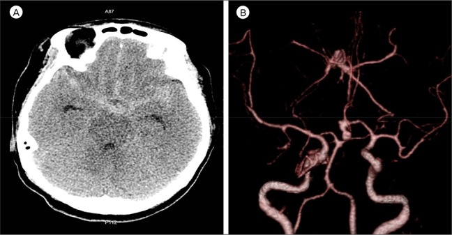
Fig. 1 A 50-year-old woman reported at the emergency department with thunderclap headache and vomiting. (A) A non-enhanced brain computed tomography (CT) showed a subarachnoid hemorrhage in the suprasellar cistern, both sylvian and interhemispheric fissure. (B) Brain angiography CT revealed an aneurysm of the anterior communicating artery. The aneurysm was left dominant, inferior in direction, and multi-lobulated.
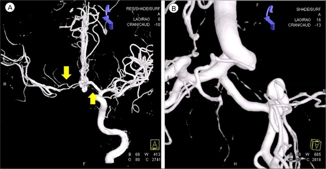
Fig. 2 A transfemoral cerebral angiography was performed pre operatively. (A, B) TThe recurrent artery of Heubner (RAH) (arrows) originated bilaterally from the junction of the anterior cerebral artery and the anterior communicating artery. The mean diameter of right and left RAH was 8 mm and 4 mm, respectively.
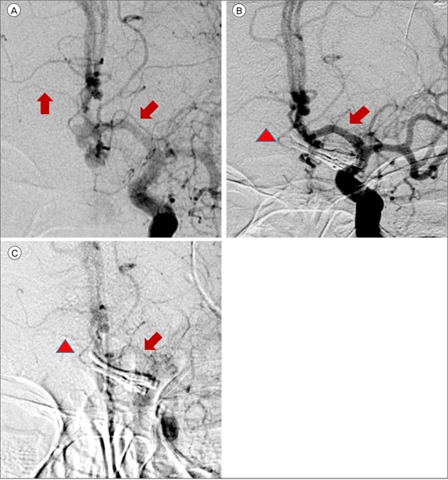
Fig. 3 (A) Both the recurrent artery of Heubner (RAH) (arrows) were evidently visible on the pre-operative transfemoral cerebral angiography (TFCA). (B) On the TFCA performed 6 days postoperatively before chemical angioplasty, a blurred right RAH (arrowhead) was seen due to decreased perfusion. The left RAH (arrow) was not visible. (C) On the TFCA performed after chemical angioplasty, the perfusion of right RAH (arrowhead) improved and left RAH (arrow) was visible.
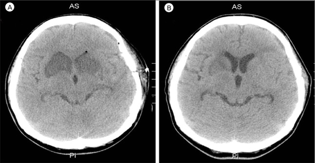
Fig. 4 (A) A 7-day postoperative follow-up brain computed tomography (CT) showed a newly developed low density lesion in the recurrent artery of Heubner (RAH) territory bilaterally, caudate nucleus, anterior portion of basal ganglia, and internal capsule. (B) The 14-day postoperative follow-up brain CT showed no progression of the low density lesion. The low density lesion in both of the RAH territory was blurred.
Reference
-
1. Berman SA, Hayman LA, Hinck VC. Correlation of CT cerebral vascular territories with function: I. Anterior cerebral artery. AJR Am J Roentgenol. 1980; 8. 135(2):253–257. PMID: 6773322.
Article2. Caplan LR, Schmahmann JD, Kase CS, Feldmann E, Baquis G, Greenberg JP, et al. Caudate infarcts. Arch Neurol. 1990; 2. 47(2):133–143. PMID: 2405818.
Article3. Critchley M. Syndromes of the anterior cerebral artery. Proc R Soc Med. 1930; 3. 23(5):630–632. PMID: 19987442.
Article4. El Falougy H, Selmeciova P, Kubikova E, Haviarová Z. The variable origin of the recurrent artery of Heubner: an anatomical and morphometric study. Biomed Res Int. 2013; 2013:873434. PMID: 23936853.
Article5. Fisher CM, Kistler JP, Davis JM. Relation of cerebral vasospasm to subarachnoid hemorrhage visualized by computerized tomographic scanning. Neurosurgery. 1980; 1. 6(1):1–9. PMID: 7354892.
Article6. Friedman JA, Goerss SJ, Meyer FB, Piepgras DG, Pichelmann MA, McIver JI, et al. Volumetric quantification of Fisher Grade 3 aneurysmal subarachnoid hemorrhage: a novel method to predict symptomatic vasospasm on admission computerized tomography scans. J Neurosurg. 2002; 8. 97(2):401–407. PMID: 12186469.7. Gomes F, Dujovny M, Umansky F, Ausman JI, Diaz FG, Ray WJ, et al. Microsurgical anatomy of the recurrent artery of Heubner. J Neurosurg. 1984; 1. 60(1):130–139. PMID: 6689705.
Article8. Kolias AG, Sen J, Belli A. Pathogenesis of cerebral vasospasm following aneurysmal subarachnoid hemorrhage: putative mechanisms and novel approaches. J Neurosci Res. 2009; 1. 87(1):1–11. PMID: 18709660.
Article9. Loukas M, Louis RG Jr, Childs RS. Anatomical examination of the recurrent artery of Heubner. Clin Anat. 2006; 1. 19(1):25–31. PMID: 16287124.
Article10. Miller SP, O'Gorman AM, Shevell MI. Recurrent artery of Heubner infarction in infancy. Dev Med Child Neurol. 2000; 5. 42(5):344–346. PMID: 10855656.
Article11. Mizuta H, Motomura N. Memory dysfunction in caudate infarction caused by Heubner's recurring artery occlusion. Brain Cogn. 2006; 7. 61(2):133–138. PMID: 16510225.
Article12. Sasaki T, Kodama N, Matsumoto M, Suzuki K, Konno Y, Sakuma J, et al. Blood flow disturbance in perforating arteries attributable to aneurysm surgery. J Neurosurg. 2007; 7. 107(1):60–67. PMID: 17639875.
Article13. Schmidt JM, Wartenberg KE, Fernandez A, Claassen J, Rincon F, Ostapkovich ND, et al. Frequency and clinical impact of asymptomatic cerebral infarction due to vasospasm after subarachnoid hemorrhage. J Neurosurg. 2008; 12. 109(6):1052–1059. PMID: 19035719.
Article14. Vergouwen MD, Ilodigwe D, Macdonald RL. Cerebral infarction after subarachnoid hemorrhage contributes to poor outcome by vasospasm-dependent and -independent effects. Stroke. 2011; 4. 42(4):924–929. PMID: 21311062.
Article15. Weidauer S, Lanfermann H, Raabe A, Zanella F, Seifert V, Beck J. Impairment of cerebral perfusion and infarct patterns attributable to vasospasm after aneurysmal subarachnoid hemorrhage: a prospective MRI and DSA study. Stroke. 2007; 6. 38(6):1831–1836. PMID: 17446425.
- Full Text Links
-
- Actions
-
Cited
- CITED
-
- Close
- Share
-
- Similar articles
-
- Microsurgical anatomy of the Anterior Cerebral-anterior Communicating Artery
- An Unruptured Anterior Communicating Artery Aneurysm with Bilateral Infraoptic Anterior Cerebral Arteries. Case Report and Review of the Literature
- Ruptured proximal anterior cerebral artery (A1) aneurysm located at an anomalous branching of the fronto-orbital artery--a case report
- Microsurgical Anatomy of the Proximal Anterior Cerebral Artery and Anterior Communicating Artery
- True Posterior Communicating Artery Aneurysm
