Posterior Vitreous Structures Evaluated by Swept-source Optical Coherence Tomography with En Face Imaging
- Affiliations
-
- 1Department of Ophthalmology, Haeundae Paik Hospital, Inje University College of Medicine, Busan, Korea. pky0402@naver.com
- KMID: 2422067
- DOI: http://doi.org/10.3341/kjo.2017.0123
Abstract
- PURPOSE
To evaluate posterior vitreous structures using swept-source (SS) optical coherence tomography (OCT) with en face imaging.
METHODS
We retrospectively reviewed OCT images of healthy individuals who did not have intra-ocular disease. We obtained high-definition horizontal and vertical line scans crossing the fovea and 3D scans using SS-OCT, with the 3D scan centered between the fovea and the optic-nerve head. An enhanced vitreous visualization function was used to highlight vitreous structures. En face mode was used to measure the area of Martegiani (AM) and bursa premacularis (BP). We performed all measurements using a built-in function of the viewing software.
RESULTS
We enrolled 24 eyes from 12 healthy individuals. The mean patient age was 28.7 ± 4.6 years (range, 24 to 39 years). The mean AM and BP areas were 5.73 ± 0.88 and 18.76 ± 6.95 mm2, respectively. In en face imaging, AM shape was most frequently a vertical oval (18 / 22, 81.8%), while the predominant BP shape was round (16 / 20, 80.0%). AM was in contact with the optic disc, either at the temporal-disc margin (13 eyes, 59.1%) or the nasal optic-disc margin (9 eyes, 40.9%).
CONCLUSIONS
Posterior vitreous structures, such as AM and BP, were readily visualized using en face imaging with SS-OCT. Investigating normal vitreous configuration might help in understanding changes in vitreous structures associated with retinal pathology.
Keyword
MeSH Terms
Figure
-

Fig. 1 (A) Twelve-millimetre horizontal line scan centered at the fovea: the enhanced vitreous visualization function was turned on, and the vitreous structure is highlighted. Horizontal and vertical arrows indicate width and central thickness of bursa premacularis, respectively. (B) Twelve-millimetre vertical line scan. (C) En face image generated from a 12 × 9-mm 3D cube scan.
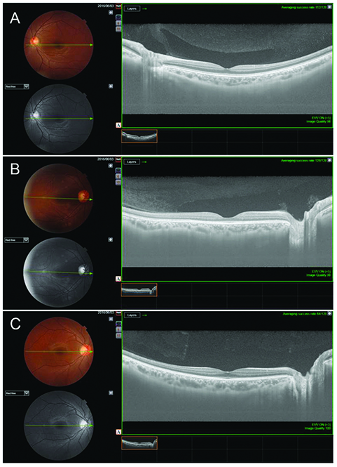
Fig. 2 Representative horizontal cross section images of bursa premacularis. (A) Boat shape, (B) wedge shape, and (C) irregular shape.

Fig. 3 Spearman correlation analysis of the optic disc area and the area of Martegiani.
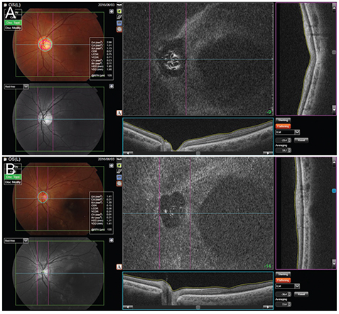
Fig. 4 Spatial relationship between optic disc margin and area of Martegiani (AM) margin. (A) The AM margin shares its temporal border with the optic disc. (B) The AM margin shares its nasal border with the optic disc.
Reference
-
1. Sebag J. Ageing of the vitreous. Eye (Lond). 1987; 1:254–262.
Article2. Sebag J. Anatomy and pathology of the vitreo-retinal interface. Eye (Lond). 1992; 6:541–552.
Article3. Duker JS, Kaiser PK, Binder S, et al. The International Vitreomacular Traction Study Group classification of vitreomacular adhesion, traction, and macular hole. Ophthalmology. 2013; 120:2611–2619.
Article4. Sebag J. Anomalous posterior vitreous detachment: a unifying concept in vitreo-retinal disease. Graefes Arch Clin Exp Ophthalmol. 2004; 242:690–698.
Article5. Itakura H, Kishi S, Li D, Akiyama H. Observation of posterior precortical vitreous pocket using swept-source optical coherence tomography. Invest Ophthalmol Vis Sci. 2013; 54:3102–3107.
Article6. Kim YC, Harasawa M, Salcedo-Villanueva G, et al. Enhanced high-density line spectral-domain optical coherence tomography imaging of the vitreoretinal interface: description of selected cases. Semin Ophthalmol. 2016; 31:559–566.
Article7. Liu JJ, Witkin AJ, Adhi M, et al. Enhanced vitreous imaging in healthy eyes using swept source optical coherence tomography. PLoS One. 2014; 9:e102950.
Article8. Park KA, Oh SY. Posterior precortical vitreous pocket in children. Curr Eye Res. 2015; 40:1034–1039.
Article9. Schaal KB, Pang CE, Pozzoni MC, Engelbert M. The premacular bursa's shape revealed in vivo by swept-source optical coherence tomography. Ophthalmology. 2014; 121:1020–1028.10. Worst JG. Cisternal systems of the fully developed vitreous body in the young adult. Trans Ophthalmol Soc U K. 1977; 97:550–554.11. Kishi S, Shimizu K. Posterior precortical vitreous pocket. Arch Ophthalmol. 1990; 108:979–982.
Article12. Spaide RF, Koizumi H, Pozzoni MC. Enhanced depth imaging spectral-domain optical coherence tomography. Am J Ophthalmol. 2008; 146:496–500.
Article13. Itakura H, Kishi S. Aging changes of vitreomacular interface. Retina. 2011; 31:1400–1404.
Article14. Itakura H, Kishi S, Li D, Akiyama H. En face imaging of posterior precortical vitreous pockets using swept-source optical coherence tomography. Invest Ophthalmol Vis Sci. 2015; 56:2898–2900.
Article15. Park SH, Lee JW, Lee MJ, et al. Evaluation of the cortical vitreous using swept-source optical coherence tomography in normal eyes. J Korean Ophthalmol Soc. 2016; 57:595–600.
Article16. Itakura H, Kishi S. Alterations of posterior precortical vitreous pockets with positional changes. Retina. 2013; 33:1417–1420.
Article
- Full Text Links
-
- Actions
-
Cited
- CITED
-
- Close
- Share
-
- Similar articles
-
- Evaluation of the Cortical Vitreous Using Swept-Source Optical Coherence Tomography in Normal Eyes
- Peripheral Lattice Degeneration Imaging with Ultra-Widefield Swept-Source Optical Coherence Tomography
- Comparison of Anterior Segment Measurements Between Swept-source Optical Coherence Tomography and Schiempflug Coherence Interferometer
- Comparison of Image Quality between Swept-Source and Spectral-Domain Optical Coherence Tomography According to Ocular Media Opacity
- Flow Void Analysis Using Different Thresholding Methods on a Choriocapillaris Optical Coherence Tomography Angiography Image Complemented with a Structural En Face Image
