J Korean Soc Spine Surg.
2018 Jun;25(2):54-59. 10.4184/jkss.2018.25.2.54.
The Effect of Cervical Lordosis on Cervical Disc Degeneration in Patients with a High T1 Slope
- Affiliations
-
- 1Department of Orthopaedic Surgery, Gwangmyeong Sungae Hospital, Gwangmyeong, Korea. java5885@gmail.com
- 2Department of Orthopaedic Surgery, Sung-Ae Hospital, Seoul, Korea.
- KMID: 2421611
- DOI: http://doi.org/10.4184/jkss.2018.25.2.54
Abstract
- STUDY DESIGN: Retrospective evaluation.
OBJECTIVES
To analyze the effect of cervical lordosis on cervical disc degeneration in patients with a high T1 slope. SUMMARY OF LITERATURE REVIEW: The T1 slope is known to be a parameter that may be very useful in evaluating sagittal balance. We previously reported that a low T1 slope was a potential risk factor for cervical spondylosis, especially in the C6-7 cervical segment. However, no study has analyzed the effect of cervical lordosis in patients with a high T1 slope (>25) on cervical disc degeneration.
MATERIALS AND METHODS
Seventy-seven patients with a high T1 slope who underwent cervical spine MRI in our orthopedic clinic were enrolled. Patients were divided into 2 groups according to cervical compensation. The radiologic parameters obtained from radiography and cervical spine MRI were compared between the uncompensated group (cervical lordosis <25) and the compensated group (cervical lordosis ≥25).
RESULTS
In the uncompensated group, the average degeneration grade of each segment was 2.72 (±0.70) in C2-3, 3.00 (±0.76) in C3-4, 3.02 (±0.91) in C4-5, 3.37 (±0.95) in C5-6, and 2.95 (±0.98) in C6-7. The average degeneration grade of each segment in the compensated group was 2.38 (±0.78) in C2-3, 2.38 (±0.60) in C3-4, 2.62 (±0.60) in C4-5, 2.82 (±0.72) in C5-6, and 2.41 (±0.74) in C6-7. The degeneration grade was significantly higher in the uncompensated group than in the compensated group for all cervical segments. The risk of high-grade degeneration of C3-4 was significantly higher in the uncompensated group (odds ratio = 6.268; 95% CI, 2.232-17.601; p<.001).
CONCLUSIONS
Patients with a high T1 slope without compensation of cervical lordosis had a higher grade of degeneration in all cervical segments.
MeSH Terms
Figure
-
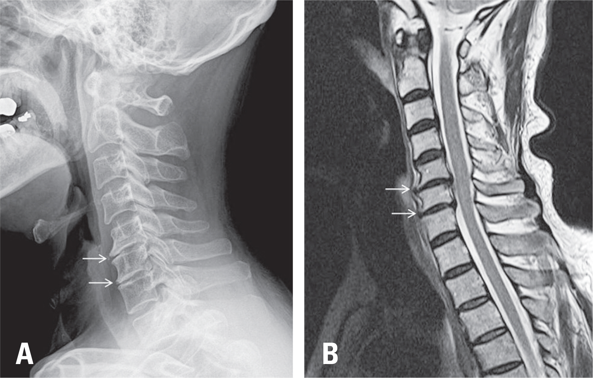
Fig 1. Radiographic example of a 55-year-old female patient without cervical compensation. (A) Upright lateral X-ray. (B) T2-weighted sagittal image on cervical magnetic resonance imaging.
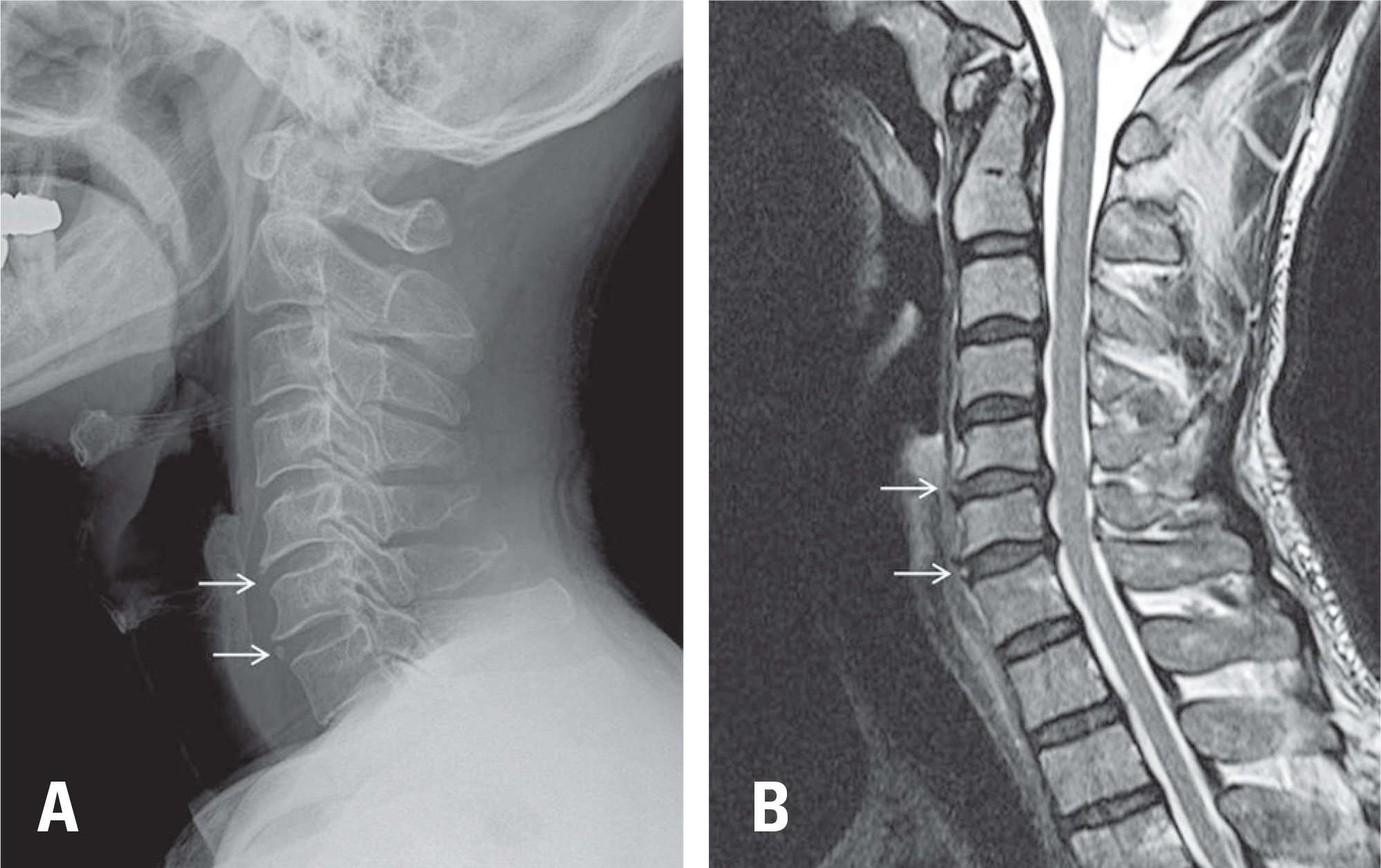
Fig 2. Radiographic example of a 51-year-old female patient with cervical compensation. (A) Upright lateral X-ray. (B) T2-weighted sagittal image on cervical magnetic resonance imaging.
Reference
-
1. Knott PT, Mardjetko SM, Techy F. The use of the T1 sagittal angle in predicting overall sagittal balance of the spine. Spine J. 2010 Nov; 10(11):994–8. DOI: 10.1016/j.spinee.2010.08.031.
Article2. Yang BS, Lee SK, Song KS, et al. The Use of T1 Sagittal Angle in Predicting Cervical Disc Degeneration. Asian Spine J. 2015 Oct; 9(5):757–61. DOI: 10.4184/asj.2015.9.5.757.
Article3. Cote P, Cassidy JD, Yong-Hing K, et al. Apophysial joint degeneration, disc degeneration, and sagittal curve of the cervical spine. Can they be measured reliably on radiographs? Spine (Phila Pa 1976). 1997 Apr 15; 22(8):859–64. DOI: DOI:10.1097/00007632-199704150-00007.4. Dai L. [Disc degeneration and cervical instability]. Zhonghua Wai Ke Za Zhi. 1999 Mar; 37(3):180–2.
Article5. DePalma AF, Rothman RH, Levitt RL, et al. The natural history of severe cervical disc degeneration. Acta Orthop Scand. 1972; 43(5):392–6. DOI: DOI:10.3109/17453677208998959.
Article6. Friedenberg ZB. Degeneration of the Cervical Disc. West J Surg Obstet Gynecol. 1964 Jul-Aug; 72:191–4.7. Gruber HE, Phillips R, Ingram JA, et al. Spontaneous age-related cervical disc degeneration in the sand rat. Clin Orthop Relat Res. 2014 Jun; 472(6):1936–42. DOI: 10.1007/s11999-014-3497-x.
Article8. Kumaresan S, Yoganandan N, Pintar FA, et al. Contribution of disc degeneration to osteophyte formation in the cervical spine: a biomechanical investigation. J Orthop Res. 2001 Sep; 19(5):977–84. DOI: 10.1016/S0736-0266 (01)00010-9.
Article9. Miyazaki M, Hymanson HJ, Morishita Y, et al. Kinematic analysis of the relationship between sagittal alignment and disc degeneration in the cervical spine. Spine (Phila Pa 1976). 2008 Nov 1; 33(23):E870–6. DOI: 10.1097/BRS.0b013e3181839733.
Article10. Sambrook PN, MacGregor AJ, Spector TD. Genetic influences on cervical and lumbar disc degeneration: a magnetic resonance imaging study in twins. Arthritis Rheum. 1999 Feb; 42(2):366–72. DOI: 10.1002/1529-0131 (199902)42: 2< 366:: AID-ANR20> 3.0.CO; 2-6.
Article11. Miyazaki M, Hong SW, Yoon SH, et al. Reliability of a magnetic resonance imaging-based grading system for cervical intervertebral disc degeneration. J Spinal Disord Tech. 2008 Jun; 21(4):288–92. DOI: 10.1097/BSD.0b013e31813c0e59.
Article12. Boden SD, McCowin PR, Davis DO, et al. Abnormal magnetic-resonance scans of the cervical spine in asymptomatic subjects. A prospective investigation. J Bone Joint Surg Am. 1990 Sep; 72(8):1178–84. DOI: DOI:10.2106/00004623-199072080-00008.
Article13. Gomez Espindola JC, Perez Viquez AF. [Cervical lordosis evaluation in asymptomatic volunteers from Navy Medical Center]. Acta Ortop Mex. 2008 Jan-Feb; 22(1):7–11.14. Wei W, Liao S, Shi S, et al. Straightened cervical lordosis causes stress concentration: a finite element model study. Australas Phys Eng Sci Med. 2013 Mar; 36(1):27–33. DOI: 10.1007/s13246-013-0182-4.
Article
- Full Text Links
-
- Actions
-
Cited
- CITED
-
- Close
- Share
-
- Similar articles
-
- The Use of T1 Sagittal Angle in Predicting Cervical Disc Degeneration
- Correlation of Cervical Disc Degeneration with Sagittal Alignments of Cervical Spine
- Relationships and Usefulness of Cervical Lateral Radiographs Compared with Whole-Spine Lateral Radiographs for Evaluating Cervical Sagittal Alignment
- Total Cervical Disc Replacement using Artificial Disc in Cervical Disc Herniations
- Early Radiological Analysis of Cervical Arthroplasty with Bryan and Mobi-C Cervical Disc Prosthesis
