Pfeiffer Syndrome Type 2 with Sporadic Fibroblast Growth Factor Receptor 2 Mutation and Coccygeal Anomaly
- Affiliations
-
- 1Department of Pediatrics, Chungnam National University Hospital, Daejeon, Korea. mychang@cnuh.co.kr
- KMID: 2421578
- DOI: http://doi.org/10.14734/PN.2018.29.3.128
Abstract
- Pfeiffer syndrome is a rare genetic disorder characterized by premature fusion of certain skull bones (craniosynostosis) and anomalies of the face and extremities. A female newborn showing ocular proptosis and typical cloverleaf skull was considered as having Pfeiffer syndrome type 2. She also had coccygeal anomaly resembling a human tail. However, the accompanying vertebral malformations are rare in Pfeiffer syndrome. Molecular genetic testing confirmed sporadic fibroblast growth factor receptor 2 mutation in this patient. Thus, molecular genetic testing should be considered for any type of Pfeiffer syndrome to obtain definite diagnosis.
MeSH Terms
-
Acrocephalosyndactylia*
Diagnosis
Exophthalmos
Extremities
Female
Fibroblast Growth Factors*
Fibroblasts*
Humans
Infant, Newborn
Molecular Biology
Receptor, Fibroblast Growth Factor, Type 2*
Receptors, Fibroblast Growth Factor*
Skull
Tail
Fibroblast Growth Factors
Receptor, Fibroblast Growth Factor, Type 2
Receptors, Fibroblast Growth Factor
Figure
-

Fig. 1 Photographs of the patient at birth. (A, B) Cloverleaf-shaped head, proptosis, hypertelorim, low-set ear, maxillary hypoplasia, and mandibular prognathism. (C) Bilateral elbow ankyloses make both arms bend into radial side with limited extension of elbows less than 90 degrees. (D) Coccygeal skin defect with human tail-like appendage.

Fig. 2 Plain radiographs of the skull and extremities. (A) Cloverleaf-shaped skull. (B) Fusion of humeroulnar, humeroradial, and radioulnar joints (black arrow). (C) The sacrococcygeal bone abruptly directed posteriorly of a lateral spine view (white arrow).
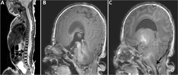
Fig. 3 Brain and spine magnetic resonance imaging. (A) The cervico-lumbar region showed kyphosis instead of lordosis, conus medllaris at L2 vertebral body (black arrow-L2 vertebral body). Posteriorly projecting coccygeal tip (white arrow). (B) Brachycephaly, hydrocephalus in the 3rd ventricle. (C) Tonsillar herniation and crowd foramen magnum (black arrow).

Fig. 4 A heterozygous mutation of c. 870G>C (p.Trp 290 Cys) in the FGFR 2 gene (arrow). Trp, trypsin; Cys, cysteine; FGFR, fibroblast growth factor receptor.
Reference
-
1. Vogels A, Fryns JP. Pfeiffer syndrome. Orphanet J Rare Dis. 2006; 1:19.
Article2. Shin CH, Yi BH, Hong HS, Lee HK, Park SJ. A case report of pfeiffer syndrome with spinal anomaly. J Korean Soc Radiol. 2009; 60:57–60.
Article3. Cohen MM Jr. Pfeiffer syndrome update, clinical subtypes, and guidelines for differential diagnosis. Am J Med Genet. 1993; 45:300–307.
Article4. Saldino RM, Steinbach HL, Epstein CJ. Familial acrocephalosyndactyly (pfeiffer syndrome). Am J Roentgenol Radium Ther Nucl Med. 1972; 116:609–622.
Article5. Lee MY, Jeon GW, Jung JM, Sin JB. A case of pfeiffer syndrome with c833_ 834GC>TG (Cys278Leu) mutation in the FGFR2 gene. Korean J Pediatr. 2010; 53:774–777.6. Chung J, Park DH, Yoon SH. Monoblock craniofacial internal distraction in a child with pfeiffer syndrome: a case report. J Korean Med Sci. 2008; 23:342–346.
Article7. Park MS, Yoo JE, Chung J, Yoon SH. A case of pfeiffer syndrome. J Korean Med Sci. 2006; 21:374–378.
Article8. Fearon JA, Rhodes J. Pfeiffer syndrome: a treatment evaluation. Plast Reconstr Surg. 2009; 123:1560–1569.
Article9. Nazzaro A, Della Monica M, Lonardo F, Di Blasi A, Baffico M, Baldi M, et al. Prenatal ultrasound diagnosis of a case of pfeiffer syndrome without cloverleaf skull and review of the literature. Prenat Diagn. 2004; 24:918–922.
Article10. Greig AV, Wagner J, Warren SM, Grayson B, McCarthy JG. Pfeiffer syndrome: analysis of a clinical series and development of a classification system. J Craniofac Surg. 2013; 24:204–215.11. Fitzgerald O'Connor EJ, Marucci DD, Jeelani NO, Witherow H, Richards R, Dunaway DJ, et al. Ocular advancement in monobloc distraction. Plast Reconstr Surg. 2009; 123:1570–1577.12. Sarkar S, Petiot A, Copp A, Ferretti P, Thorogood P. FGF2 promotes skeletogenic differentiation of cranial neural crest cells. Development. 2001; 128:2143–2152.
Article13. OMIM and Online Mendelian Inheritance in Man. #101600 Pfeiffer syndrome. accessed on 1 Aug, 2015. Available at http://omim.org/entry/101600.14. Rossi M, Jones RL, Norbury G, Bloch-Zupan A, Winter RM. The appearance of the feet in pfeiffer syndrome caused by FGFR1 P252R mutation. Clin Dysmorphol. 2003; 12:269–274.
Article15. Tartaglia M, Valeri S, Velardi F, Di Rocco C, Battaglia PA. Trp290Cys mutation in exon IIIa of the fibroblast growth factor receptor 2 (FGFR2) gene is associated with pfeiffer syndrome. Hum Genet. 1997; 99:602–606.
Article16. Schaefer F, Anderson C, Can B, Say B. Novel mutation in the FGFR2 gene at the same codon as the crouzon syndrome mutations in a severe pfeiffer syndrome type 2 case. Am J Med Genet. 1998; 75:252–255.
Article17. Gonzales M, Heuertz S, Martinovic J, Delahaye S, Bazin A, Loget P, et al. Vertebral anomalies and cartilaginous tracheal sleeve in three patients with Pfeiffer syndrome carrying the S351C FGFR2 mutation. Clin Genet. 2005; 68:179–181.18. Mukhopadhyay B, Shukla RM, Mukhopadhyay M, Mandal KC, Haldar P, Benare A. Spectrum of human tails: a report of six cases. J Indian Assoc Pediatr Surg. 2012; 17:23–25.
Article
- Full Text Links
-
- Actions
-
Cited
- CITED
-
- Close
- Share
-
- Similar articles
-
- A case of Pfeiffer syndrome with c833_834GC>TG (Cys278Leu) mutation in the FGFR2 gene
- A Case of FGFR2 Exon lllc Mutation in Crouzon Syndrome
- Apert and Pfeiffer Syndromes: A Report of Two Cases
- Mutation at exon 10 of the fibroblast growth factor receptor 3 (FGFR3) in a fetus with thanatophoric dysplasia type I (TDI)
- A case of Apert's Syndrome(Acrocophalosyndactyly) with Fibroblast Growth Factor Receptor 2 Exon IIIa Mutation
