J Korean Soc Radiol.
2018 Oct;79(4):233-236. 10.3348/jksr.2018.79.4.233.
Popliteal Artery Agenesis Detected by CT Angiography
- Affiliations
-
- 1Department of Radiology, Kosin University College of Medicine, Gospel Hospital, Busan, Korea. gsjung240@gmail.com
- 2Department of Internal Medicine, Kosin University College of Medicine, Gospel Hospital, Busan, Korea.
- KMID: 2421227
- DOI: http://doi.org/10.3348/jksr.2018.79.4.233
Abstract
- Variations in the popliteal artery and its tibial branches are common and have assumed greater importance with the technological advances that have made below-knee interventional revascularization feasible. The authors report a rare anatomic variant of popliteal artery agenesis, describing the CT angiographic findings with emphasis on the differential point from the occlusive disease and reviewing the embryologic development of the lower extremity vasculature.
Figure
-
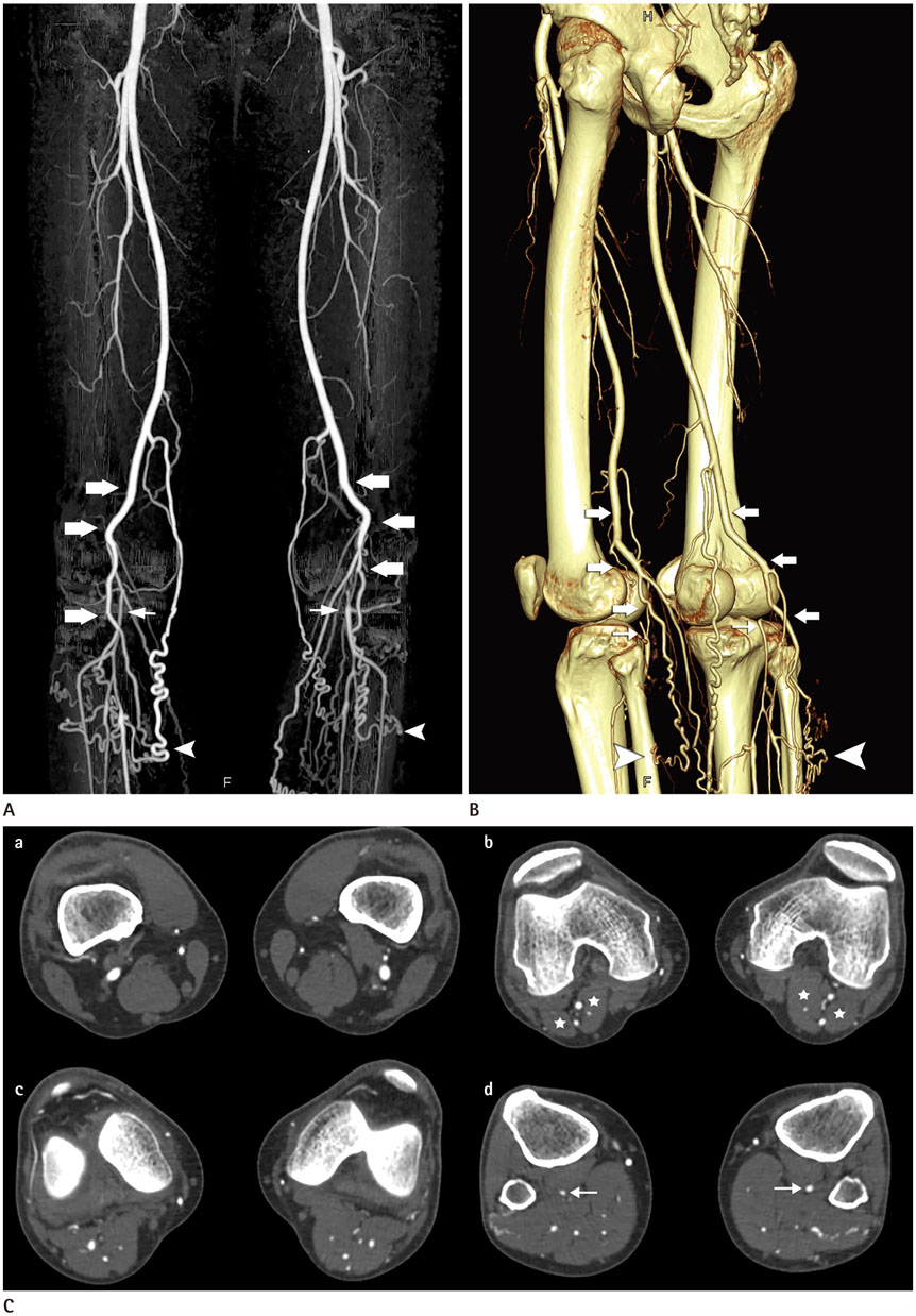
Fig. 1 CT angiographic images of a 48-year-old man with bilateral popliteal artery agenesis. A, B. Maximum intensity projection (A) and volume rendering (B) images in posterolateral projection show bilateral popliteal arteries ended gradually as prominent muscular branches (thick arrows). Notice abundant collateral arteries (arrowheads) reconstituting a distal portion of a popliteal artery (thin arrows) on both sides. C. Axial images show (a) normal P1 segment, (b, c) absent popliteal artery above the knee, and (d) reconstituted P3 vessel (arrows) on both sides. Gastrocnemius muscles (asterisks) are normal.
Reference
-
1. Kim D, Orron DE, Skillman JJ. Surgical significance of popliteal arterial variants. a unified angiographic classification. Ann Surg. 1989; 210:776–781.2. Kelly AM, Cronin P, Hussain HK, Londy FJ, Chepeha DB, Carlos RC. Preoperative MR angiography in free fibula flap transfer for head and neck cancer: clinical application and influence on surgical decision making. AJR Am J Roentgenol. 2007; 188:268–274.
Article3. Kil SW, Jung GS. Anatomical variations of the popliteal artery and its tibial branches: analysis in 1242 extremities. Cardiovasc Intervent Radiol. 2009; 32:233–240.
Article4. Neville RF Jr, Franco CD, Anderson RJ, Padberg FT Jr, Hobson RW 2nd. Popliteal artery agenesis: a new anatomic variant. J Vasc Surg. 1990; 12:573–576.
Article5. Mauro MA, Jaques PF, Moore M. The popliteal artery and its branches: embryologic basis of normal and variant anatomy. AJR Am J Roentgenol. 1988; 150:435–437.
Article6. Waugh JR, Sacharias N. Arteriographic complications in the DSA era. Radiology. 1992; 182:243–246.
Article7. Sun Z. Diagnostic accuracy of multislice CT angiography in peripheral arterial disease. J Vasc Interv Radiol. 2006; 17:1915–1921.
Article8. Zhong H, Gan J, Zhao Y, Xu Z, Liu C, Shao G, et al. Role of CT angiography in the diagnosis and treatment of popliteal vascular entrapment syndrome. AJR Am J Roentgenol. 2011; 197:W1147–W1154.
Article9. Wright LB, Matchett WJ, Cruz CP, James CA, Culp WC, Eidt JF, et al. Popliteal artery disease: diagnosis and treatment. Radiographics. 2004; 24:467–479.
Article10. Foley WD, Stonely T. CT angiography of the lower extremities. Radiol Clin North Am. 2010; 48:367–396.
Article
- Full Text Links
-
- Actions
-
Cited
- CITED
-
- Close
- Share
-
- Similar articles
-
- Cystic Adventitial Disease of the Popliteal Artery as Demonstrated by MDCT Angiography: A Case Report
- Adventitial Cystic Disease of Popliteal Artery
- Agenesis of Right Internal Carotid Artery Associated with Intracranial Aneurysm: Case Report
- Total Popliteal Artery Occlusion after Stent Insertion Diagnosed as Popliteal Artery Entrapment Syndrome
- Delayed Onset of the Popliteal Artery Pseudoaneurysm Following Medial Opening Wedge High Tibial Osteotomy
