Evaluation of gallbladder and common bile duct size and appearance by computed tomography in dogs
- Affiliations
-
- 1Department of Veterinary Medical Imaging, College of Veterinary Medicine, Chungnam National University, Daejeon 34134, Korea. hjchoi@cnu.ac.kr
- KMID: 2420934
- DOI: http://doi.org/10.4142/jvs.2018.19.5.653
Abstract
- The feasibility of using computed tomography (CT) to identify the common bile duct (CBD) and comparison with ultrasonography (US) results were evaluated in normal beagle dogs and dogs without hepatobiliary and pancreatic diseases. In addition, CBD diameters were obtained from CT at the level of the porta hepatis and the duodenal papilla level in dogs with underlying diseases that may cause cholestasis. US is a useful modality in the estimation of gallbladder volume because ejection fraction and CBD diameter from US were not significantly different from those of CT. The normal biliary tract was visible on CT images in 68% of the normal dog group. CBD diameter was not over 3 mm and 3.5 mm at the porta hepatis and duodenal papilla levels, respectively in normal dogs weighing less than 15 kg. Dogs suspected to have cholestasis associated with hepatobiliary or pancreatic diseases had significantly larger CBD than that in normal dogs.
MeSH Terms
Figure
-
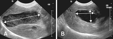
Fig. 1 Ultrasonographic images of gallbladder (GB) in longitudinal (A) and transverse (B) planes. Maximum length (A; double arrow), width (B; double arrow) and depth (B; dashed double arrow) were measured to evaluate GB volume.
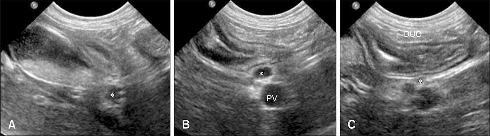
Fig. 2 Ultrasonographic images of the bile duct in a normal beagle illustrating where the three duct diameters were measured. Cystic duct in gallbladder (GB) level (A), common bile duct (CBD) at the porta hepatis level (B), and CBD at the duodenal papilla level (C) on transverse (A) and sagittal images (B and C). Diameter of bile duct (*) is measured as the maximum length perpendicular to long axis of the duct. PV, portal vein; DUO, duodenum.
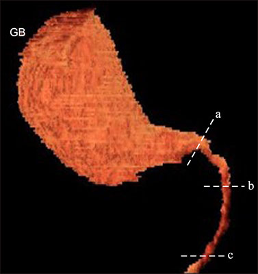
Fig. 3 Representative reconstructed three-dimensional (3D) volume rendered image of gallbladder (GB). The GB volume was measured through the application of 3D volume rendering. Bile duct diameters were measured at three measurement sites: cystic duct at the GB level (a), common bile duct (CBD) at the porta hepatis level (b), and CBD at the duodenal papilla level (c).
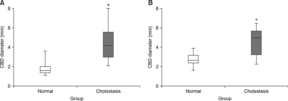
Fig. 4 Box and whisker plots of common bile duct (CBD) diameter in the normal and cholestasis groups. CBD diameters were measured at the porta hepatis (A) and duodenal papilla (B) levels. CBD diameter of the cholestasis group was significantly higher than that in the normal group (*p < 0.05).
Reference
-
1. Atalan G, Barr FJ, Holt PE. Estimation of the volume of the gall bladder of 32 dogs from linear ultrasonographic measurements. Vet Rec. 2007; 160:118–122.
Article2. Brook OR, Suissa A, Khamaysi I, Koren D, Gaitini D. Difference of CBD width on US vs. ERCP. Abdom Imaging. 2007; 32:652–656.
Article3. Co CS, Shea WJ Jr, Goldberg HI. Evaluation of common bile duct diameter using high resolution computed tomography. J Comput Assist Tomogr. 1986; 10:424–427.4. Dolmatch BL, Laing FC, Ferderle MP, Jeffrey RB, Cello J. AIDS-related cholangitis: radiographic findings in nine patients. Radiology. 1987; 163:313–316.
Article5. Finn-Bodner ST, Park RD, Tyler JW, Twedt DC, Curtis CR. Ultrasonographic determination, in vitro and in vivo, of canine gallbladder volume, using four volumetric formulas and stepwise-regression models. Am J Vet Res. 1993; 54:832–835.6. Foley WD, Wilson CR, Quiroz FA, Lawson TL. Demonstration of the normal extrahepatic biliary tract with computed tomography. J Comput Assist Tomogr. 1980; 4:48–52.
Article7. Gaschen L. Update on hepatobiliary imaging. Vet Clin North Am Small Anim Pract. 2009; 39:439–467.
Article8. Grand D, Horton KM, Fishman EK. CT of the gallbladder: spectrum of disease. Am J Roentgenol. 2004; 183:163–170.
Article9. Haaga JR, Boll D. Gastrointestinal imaging. In : Dogra VS, Forsting M, Gilkeson RC, Ha KH, Sundaram M, editors. CT and MRI of the Whole Body. 5th ed. Maryland: Mosby;2008. p. 1213–1806.10. Kim JE, Lee JK, Lee KT, Park DI, Hyun JG, Paik SW, Rhee JC, Choi KW, Lim JH. The clinical significance of common bile-duct dilatation in patients without biliary symptoms or causative lesions on ultrasonography. Endoscopy. 2001; 33:495–500.
Article11. Léveillé R, Biller DS, Shiroma JT. Sonographic evaluation of the common bile duct in cats. J Vet Intern Med. 1996; 10:296–299.
Article12. Rahmani V, Molazem M, Jamshidi S, Vali Y, Hanifeh M. Evaluation of gallbladder volume and contraction index with three-dimensional ultrasonography in healthy dogs. J Vet Med Sci. 2015; 77:1157–1161.
Article13. Ramstedt KL, Center SA, Randolph JF, Yeager AE, Erb HN, Warner KL. Changes in gallbladder volume in healthy dogs after food was withheld for 12 hours followed by ingestion of a meal or a meal containing erythromycin. Am J Vet Res. 2008; 69:647–651.
Article14. Romański KW, Siembieda J. Validation of the methods estimating gallbladder volume. Bull Vet Inst Pulawy. 2002; 46:95–104.15. Schulte SJ, Baron RL, Teefey SA, Rohrmann CA Jr, Freeny PC, Shuman WP, Foster MA. CT of the extrahepatic bile ducts: wall thickness and contrast enhancement in normal and abnormal ducts. Am J Roentgenol. 1990; 154:79–85.
Article16. Songür Y, Temuçin G, Sahin B. Endoscopic ultrasonography in the evaluation of dilated common bile duct. J Clin Gastroenterol. 2001; 33:302–305.
Article17. Thompson SM. Biliary tract surgery in the dog: a review. J Small Anim Pract. 1981; 22:437–450.
Article18. Tsukagoshi T, Ohno K, Tsukamoto A, Fukushima K, Takahashi M, Nakashima K, Fujino Y, Tsujimoto H. Decreased gallbladder emptying in dogs with biliary sludge or gallbladder mucocele. Vet Radiol Ultrasound. 2012; 53:84–91.
Article19. Turner MA, Fulcher AS. The cystic duct: normal anatomy and disease processes. Radiographics. 2001; 21:3–22.
Article20. Van den Bossche I, Paepe D, Daminet S. Acute pancreatitis in dogs and cats: pathogenesis, clinical signs and clinicopathologic findings. Vlaams Diergeneeskd Tijdschr. 2010; 79:13–22.21. Yeh BM, Liu PS, Soto JA, Corvera CA, Hussain HK. MR imaging and CT of the biliary tract. RadioGraphics. 2009; 29:1669–1688.
Article22. Yoneda M, Tamasawa N, Makino I, Takebe K, Sakuraba K, Tamura T. Measurement of calcium content of gallstones by computed tomography and the relationship between gallbladder function and calcification of gallstones. Gastroenterol Jpn. 1990; 25:478–484.
Article23. Zeman RK, Taylor KJ, Rosenfield AT, Schwartz A, Gold JA. Acute experimental biliary obstruction in the dog: sonographic findings and clinical implications. Am J Roentgenol. 1981; 136:965–967.
Article
- Full Text Links
-
- Actions
-
Cited
- CITED
-
- Close
- Share
-
- Similar articles
-
- Laparoscopic cholecystectomy and common bile duct exploration for gallstone and common bile duct stone in a patient with a left-sided gallbladder: a case report
- Ultrasound-guided transhepatic computed tomography cholecystography in beagle dogs
- Measurement of the Bile Duct in Korean Normal Adult
- Anomalous Drainage of the Common Bile Duct and Pancreatic Duct into the Duodenal Bulb
- A case of limy bile accompanied with gallbladder stones and common bile duct stone
