Chonnam Med J.
2018 Sep;54(3):197-198. 10.4068/cmj.2018.54.3.197.
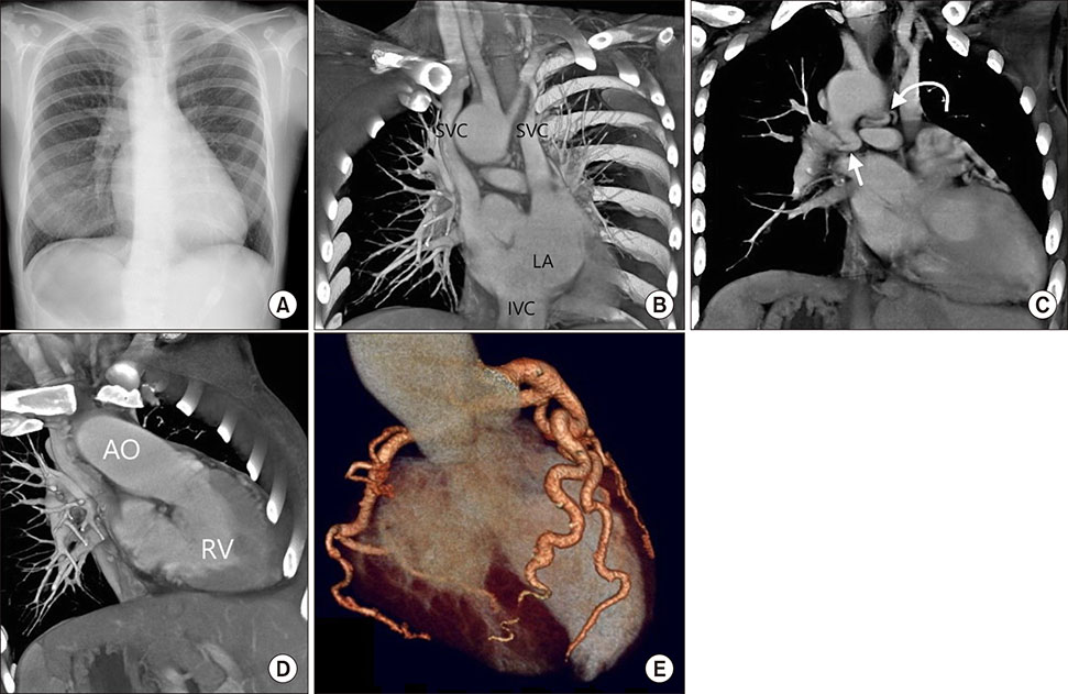
An Unusual Adult Complex Congenital Heart Disease
- Affiliations
-
- 1Division of Cardiology, Department of Internal Medicine, Chang Gung University College of Medicine, Kaohsiung, Taiwan, Republic of China. leeweichieh@yahoo.com.tw, chuasr409@hotmail.com
- 2Department of Radiology Kaohsiung Chang Gung Memorial Hospital, Chang Gung University College of Medicine, Kaohsiung, Taiwan, Republic of China.
- KMID: 2420889
- DOI: http://doi.org/10.4068/cmj.2018.54.3.197
Abstract
- No abstract available.
MeSH Terms
Figure
Reference
-
1. Presbitero P, Somerville J, Rabajoli F, Stone S, Conte MR. Corrected transposition of the great arteries without associated defects in adult patients: clinical profile and follow-up. Br Heart J. 1995; 74:57–59.
Article2. Hornung TS, Calder L. Congenitally corrected transposition of the great arteries. Heart. 2010; 96:1154–1161.
Article
- Full Text Links
- Actions
-
Cited
- CITED
-
- Close
- Share
- Similar articles
-
- Differential Diagnosis of Congenital Heart Diseases
- Echocardiographic Evaluation of Complex Congenital Heart Disease
- Growth of right ventricular outflow tract after "REV" operation in complex congenital heart disease
- Heart-Lung Transplantation in Korea
- Transitioning Adolescents with Congenital Heart Disease into Adult Health Care


