Comparison of Miniflap and Rotational Conjunctival Flap Techniques of Pterygium Accompanied by Conjunctivochalasis
- Affiliations
-
- 1Department of Ophthalmology, Dong-A University College of Medicine, Busan, Korea. wcpark@dau.ac.kr
- KMID: 2420328
- DOI: http://doi.org/10.3341/jkos.2018.59.9.810
Abstract
- PURPOSE
To investigate the differences between the recurrence and cosmesis of the miniflap technique and rotational conjunctival flap technique using redundant conjunctiva tissue in pterygium patients accompanied by conjunctivochalasis.
METHODS
This retrospective clinical study included 48 patients diagnosed with pterygium and conjunctivochalsis: 27 pterygium patients who underwent surgery using the miniflap technique and 21 pterygium patients who underwent surgery using the rotational conjunctival flap technique with redundant conjunctiva tissue. The recurrence of each operation, defined as fibrovascular invasion of the cornea, was analyzed. Redness was compared using Image J software to resolve the red color ratios with respect to the red, green, and blue colors of the pterygium excision site. The difference in the red color ratio before and after surgery was analyzed with each surgery.
RESULTS
For patients who underwent the miniflap technique, the mean age was 66 years, the recurrence rate was 5.1%, the mean follow-up period was 12 months, and the average recurrence was 5 months after surgery. For patients who underwent the rotational conjunctival flap technique using redundant conjunctiva tissue, the mean age was 62 years, the recurrence rate was 5.9%, the mean follow-up period was 20 months, and the average recurrence was 5 months after surgery. The ratio of the red color after surgery decreased 5.3% in the miniflap technique group and 6.1% in the rotational conjunctival flap technique group. Between the two groups, there was no significant difference in the recurrence or redness after surgery.
CONCLUSIONS
The rotational conjunctival flap technique using redundant conjunctiva in patients with pterygium and conjunctivochalasis was useful for reducing recurrence and cosmesis.
MeSH Terms
Figure
-
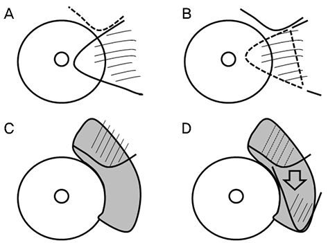
Figure 1 The surgical procedure of modified miniflap technique using amniotic membrane transplantation. (A) A small conjunctival flap was made (dotted line) before fibrovascular tissues of pterygium (thin lines) excision. (B) The pterygium head and body were excised, and subconjunctival fibrovascular tissue was removed (area of thin lines surrounded with dotted line). (C) Amniotic membrane is placed over exposed sclera, and was sutured (dark area), and small conjunctival flap was placed over amniotic membrane transplantation area (area of thin lines surrounded with thick line). (D) The small conjunctival flap was transposed to pterygium excision area over amniotic membrane transplantation site (area of thin lines).
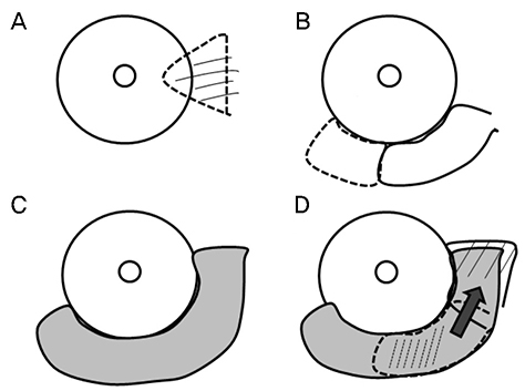
Figure 2 The surgical procedure of rotational conjunctival flap technique with redundant conjunctiva tissue. (A) The pterygium head and body were excised (area of thin lines surrounded with dotted line). (B) The redundant conjunctiva of temporal area (area of dotted line) was excised. Conjunctival flap was made with remained redundant conjunctiva (thick line). (C) Amniotic membrane is placed over exposed sclera of pterygium excision, conjunctival flap, and redundant conjunctiva excision site and was sutured (dark area). (D) The conjunctival flap (area of dotted line) was transposed to pterygium excision area (area of thin lines) over amniotic membrane transplantation site (dark area).

Figure 3 The rotational conjunctival flap technique of pterygium surgery accompanied with conjunctival chalasis. (A) Before surgery, dotted line shows conjunctival chalasis. (B) After pterygium excision, temporal area of redundant conjunctiva was excised. And conjunctival flap was made with remained redundant conjunctiva. After conjunctival flap was made, amniotic membrane transplantation was done on the exposed sclera of pterygium excision, conjunctival flap, redundant conjunctiva excision site. And conjunctival flap was transposed to pterygium excision site (dotted line).
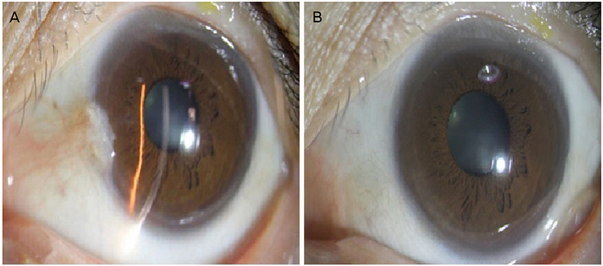
Figure 4 Anterior segment photographs of miniflap technique. (A) Anterior segment photography before miniflap technique. (B) Postoperative 6 months in patients received miniflap technique.
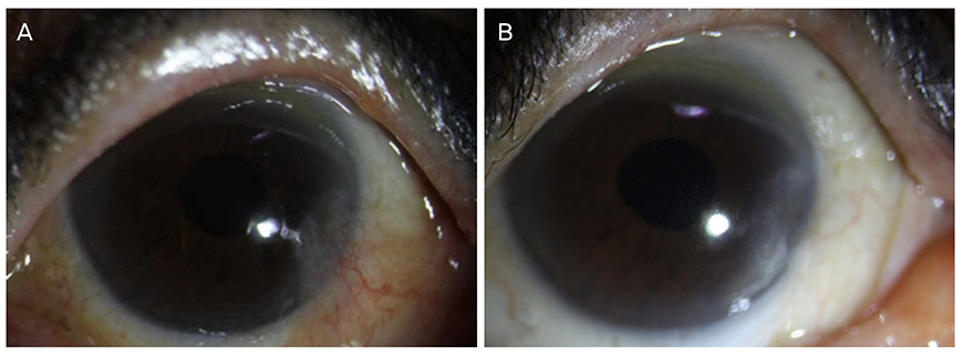
Figure 5 Anterior segment photographs of conjunctival flap technique. (A) Anterior segment photography before conjunctival flap technique accompanied with conjunctival chalasis. (B) Postoperative 6 months in patients received rotational conjunctival flap technique using redundant conjunctiva.
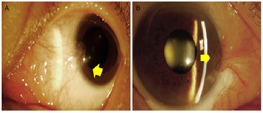
Figure 6 Anterior segment photographs of pterygium recurrence. (A) Anterior segment photopraphy shows recurrence after pterygium surgery. Recurrence after pterygium surgery was defined when fibrovascular tissue invaded cornea (yellow arrow). (B) Anterior segment photography shows corneal neovascularization after pterygium surgery (yellow arrow).

Figure 7 The Photographs show red-green-blue (RGB) analysis of Image J program. RGB analysis was applied on the operation site except the cornea area (yellow lined area).
Reference
-
1. Janson BJ, Sikder S. Surgical management of pterygium. Ocul Surf. 2014; 12:112–119.
Article2. Liu D. Conjunctivochalasis. A cause of tearing and its management. Ophthalmic Plast Reconstr Surg. 1986; 2:25–28.3. Wang Y, Dogru M, Matsumoto Y, et al. The impact of nasal conjunctivochalasis on tear functions and ocular surface findings. Am J Ophthalmol. 2007; 144:930–937.
Article4. Arenas E, Muñoz D. A new surgical approach for the treatment of conjunctivochalasis: reduction of the conjunctival fold with bipolar electrocautery forceps. Scientific World Journal. 2016; 2016:6589751. DOI: 10.1155/2016/6589751.
Article5. Qiu W, Zhang M, Xu T, et al. Evaluation of the effects of conjunctivochalasis excision on tear stability and contrast sensitivity. Scientific reports. 2016; 6:37570.
Article6. Anbesse DH, Kassa T, Kefyalew B, et al. Prevalence and associated factors of pterygium among adults living in Gondar city, Northwest Ethiopia. PLoS One. 2017; 12:e0174450. eCollection 2017. DOI: 10.1371/journal.pone.0174450.
Article7. Gumus K, Pflugfelder SC. Increasing prevalence and severity of conjunctivochalasis with aging detected by anterior segment optical coherence tomography. Am J Ophthalmol. 2013; 155:238–242.e2.
Article8. Tong L, Lan W, Shim HS, Hou A. Conjunctivochalasis is the precursor to pterygium. Med Hypotheses. 2013; 81:927–930.
Article9. Solomon A, Li DQ, Lee SB, Tseng SC. Regulation of collagenase, stromelysin, and urokinase-type plasminogen activator in primary pterygium body fibroblasts by inflammatory cytokines. Invest Ophthalmol Vis Sci. 2000; 41:2154–2163.10. Ward SK, Wakamatsu TH, Dogru M, et al. The role of oxidative stress and inflammation in conjunctivochalasis. Invest Ophthalmol Vis Sci. 2010; 51:1994–2002.
Article11. Yu XY, Jian ZY, Wu W, Lu XH. Simultaneous treatment of pterygium complicated with conjunctivochalasis: analysis of pterygium excision and conjunctival autotransplantation combined with sclera fixation. BMC Ophthalmology. 2015; 15:100. DOI: 10.1186/s12886-015-0057-4.
Article12. Suzuki H, Shiwa T, Oharazawa H, et al. Simultaneous treatment of pterygium and temporal conjunctivochalasis. J Nippon Med Sch. 2013; 80:74–77.
Article13. Meller D, Tseng SC. Conjunctivochalasis: literature review and possible pathophysiology. Surv Ophthalmol. 1998; 43:225–232.14. Zhang X, Li Q, Zou H, et al. Assessing the severity of conjunctivochalasis in a senile population: a community-based epidemiology study in Shanghai, China. BMC Public Health. 2011; 11:198. DOI: 10.1186/1471-2458-11-198.
Article15. Cornelius CR. Recurrence rate and complications of pterygium extended removal followed by extended conjunctival transplant. Cornea. 2017; 36:101–103.
Article16. Akura J, Kaneda S, Matsuura K, et al. Measures for preventing recurrence after pterygium surgery. Cornea. 2001; 20:703–707.
Article17. Cho JW, Chung SH, Seo KY, Kim EK. Conjunctival mini-flap technique and conjunctival autotransplantation in pterygium surgery. J Korean Ophthalmol Soc. 2005; 46:1471–1477.18. Park SY, Han KE, Seo KR. Recurrence after modified mini-flap technique for pterygium surgery. J Korean Ophthalmol Soc. 2012; 53:1419–1424.
Article19. Alpay A, Uğurbaş SH, Erdoğan B. Comparing techniques for pterygium surgery. Clin Ophthalmol. 2009; 3:69–74.20. Hirst LW. The Treatment of pterygium. Surv Ophthalmol. 2003; 48:145–180.
Article21. Hirst LW. Recurrent pterygium surgery using pterygium extended removal followed by extended conjunctival transplant: recurrence rate and cosmesis. Ophthalmology. 2009; 116:1278–1286.
Article22. Prabhasawat P, Barton K, Burkett G, Tseng SC. Comparison of conjunctival autografts, amniotic membrane grafts, and primary closure for pterygium excision. Ophthalmology. 1997; 104:974–985.
Article23. Küçükerdönmez C, Akova Y, Altönörs DD. Comparison of conjunctival autograft with amniotic membrane transplantation for pterygium surgery: surgical and cosmetic outcome. Cornea. 2007; 26:407–413.24. Hirst LW. Cosmesis after pterygium extended removalfollowed by extended conjunctival transplant as assessed by a new, web-based grading system. Ophthalmology. 2011; 118:1739–1746.25. Meller D, Maskin SL, Pires RT, Tseng SC. Amniotic membrane transplantation for symptomatic conjunctivochalasis refractory to medical treatments. Cornea. 2000; 19:796–803.
Article26. Fernández-Hortelano A, Moreno-Montañés J, Heras-Mulero H, Sadaba-Echarri LM. Amniotic membrane transplantation with fibrin glue as treatment of refractory conjunctivochalasis. Arch Soc Esp Oftalmol. 2007; 82:571–574.27. Kheirkhah A, Casas V, Blanco G, et al. Amniotic membrane transplantation with fibrin glue for conjunctivochalasis. Am J Ophthalmol. 2007; 144:311–313.
Article28. Georgiadis NS, Terzidou CD. Epiphora caused by conjunctivochalasis: treatment with transplantation of preserved human amniotic membrane. Cornea. 2001; 20:619–621.29. Kim SH, Oh JH, Do JR, et al. A Comparison of anchored conjunctival rotation flap and conjunctival autograft techniques in pterygium surgery. Cornea. 2013; 32:1578–1581.
Article
- Full Text Links
-
- Actions
-
Cited
- CITED
-
- Close
- Share
-
- Similar articles
-
- Conjunctival Mini-flap Technique and Conjunctival Autotransplantation in Pterygium Surgery
- Effect of Inferior Conjunctival Transposition Flap Surgery for Primary Pterygium
- Autogenous Temporalis Fascia Grafting and Conjunctival Flap Transposition in Scleromalacia after Pterygium Excision
- A Case of Conjunctival Autotransplantation Using Conjunctival Flap of Pterygium in Treating Corneal Ulcer Perforation
- Surgical Outcome of Primary Pterygium Excision with Conjunctival Autograft
