First Case of Plasma Cell Myeloma With Brown Tumor Features Unrelated to Hyperparathyroidism
- Affiliations
-
- 1Department of Laboratory Medicine and Genetics, Samsung Medical Center, Sungkyunkwan University School of Medicine, Seoul, Korea. sunnyhk@skku.edu
- 2Department of Medicine, Samsung Medical Center, Sungkyunkwan University School of Medicine, Seoul, Korea.
- 3Department of Laboratory Medicine, Ajou University Hospital of School of Medicine, Suwon, Korea.
- KMID: 2420278
- DOI: http://doi.org/10.3343/alm.2019.39.1.96
Abstract
- No abstract available.
Figure
-
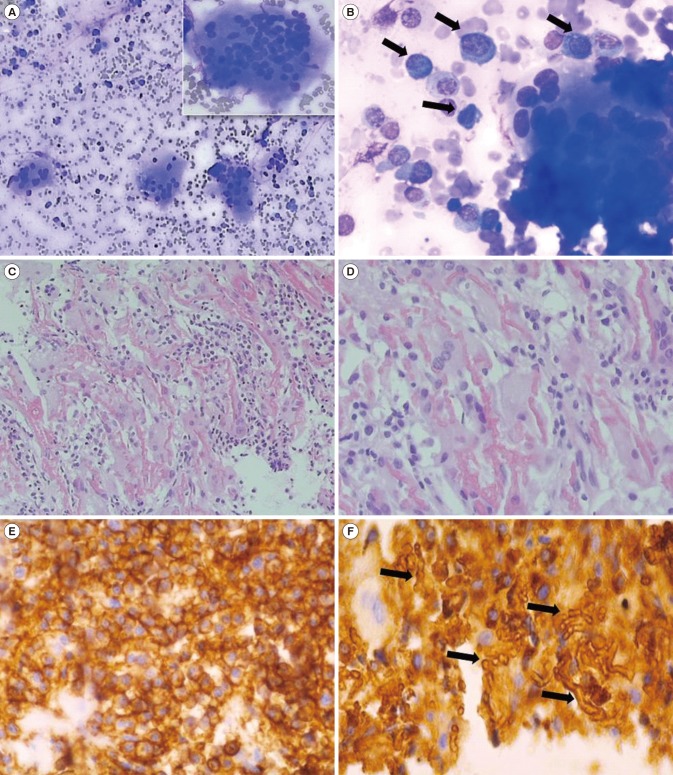
Fig. 1 Features of bone marrow aspiration and biopsy. (A, B) Bone marrow aspirate showing increased plasma cells (black arrows) with markedly increased osteoclast clusters (Wright-Giemsa stain, ×100 & ×400). A multinucleated osteoclast is magnified in right upper section of Fig. 1A. (C, D) Bone marrow biopsy with tunneling feature as well as interstitial hemorrhage and woven bone formation following trabecular destruction and increase in osteoclast proliferation (hematoxylin & eosin, ×100 & ×400). (E) Immunohistochemical staining was positive for CD138 in bone marrow biopsy. (F) Ig kappa light chain on malignant plasma cells. Interstitial hemorrhage was observed in the form of crystals (black arrows) after Ig kappa light chain staining (×400).
Reference
-
1. Mckenna RW, Kyle RA, et al. Plasma cell neoplasms. In : Swerdlow SH, Campo E, editors. WHO classification of tumours of haematopoietic and lymphoid tissues. 4th ed. Lyon: IARC;2017. p. 241–258.2. Bingham N, Reale A, Spencer A. An evidence-based approach to myeloma bone disease. Curr Hematol Malig Rep. 2017; 12:109–118. PMID: 28243849.3. Terpos E, Christoulas D, Gavriatopoulou M, Dimopoulos MA. Mechanisms of bone destruction in multiple myeloma. Eur J Cancer Care (Engl). 2017; 26.4. Bassler T, Wong ET, Brynes RK. Osteitis fibrosa cystica simulating metastatic tumor. An almost-forgotten relationship. Am J Clin Pathol. 1993; 100:697–700. PMID: 8249919.5. Ullah E, Ahmad M, Ali SA, Redhu N. Primary hyperparathyroidism having multiple brown tumors mimicking malignancy. Indian J Endocrinol Metab. 2012; 16:1040–1042. PMID: 23226663.6. Lee JH, Chung SM, Kim HS. Osteitis fibrosa cystica mistaken for malignant disease. Clin Exp Otorhinolaryngol. 2013; 6:110–113. PMID: 23799171.7. Zhang J, Wang H, Tian W, He Q, Zhu M. Brown tumor of the rib as a first presentation of primary hyperparathyroidism: report of three cases and literature review. Thorac Cancer. 2013; 4:474–478. PMID: 28920228.8. Jervis L, James M, Howe W, Richards S. Osteolytic lesions: osteitis fibrosa cystica in the setting of severe primary hyperparathyroidism. BMJ Case Rep. 2017; 2017:bcr-2017-220603.9. Bains MA, Pardoe LE, Rudin CE. Osteitis fibrosa cystica and secondary hyperparathyroidism in multiple myeloma. Br J Haematol. 2007; 136:179. PMID: 17062009.
- Full Text Links
-
- Actions
-
Cited
- CITED
-
- Close
- Share
-
- Similar articles
-
- Multiple brown tumors in a patient with primary hyperparathyroidism
- A Case of Brown Tumor of the Hard Palate in Association with Primary Hyperparathyroidism
- Brown Tumor of the Cervical Spines: A Case Report with Literature Review
- Multiple brown tumors of the jaws in primary hyperparathyroidism
- SOLITARY PLASMA CELL MYELOMA ON ANTERIOR MAXILLA: A CASE REPORT
