Ann Dermatol.
2018 Jun;30(3):331-334. 10.5021/ad.2018.30.3.331.
Dermoscopic “Landscape Painting Patterns†as a Clue for Labial Melanotic Macules: An Analysis of 80 Cases
- Affiliations
-
- 1Department of Dermatology, Pusan National University School of Medicine, Busan, Korea. drkmp@hanmail.net
- 2Biomedical Research Institute, Pusan National University Hospital, Busan, Korea.
- KMID: 2419175
- DOI: http://doi.org/10.5021/ad.2018.30.3.331
Abstract
- BACKGROUND
Labial melanotic macules (LMMs) are benign pigmented lesions that usually take the shape of flat asymmetrical macules with tan-brown to black color and variable size. Whereas the dermoscopic features of other pigmented skin lesions have been relatively well described, little is known about LMMs.
OBJECTIVE
To describe the dermoscopic features and find typical and schematic dermoscopic patterns in LMMs.
METHODS
A retrospective dermoscopic study was conducted on 80 lesions with histopathologically proved LMMs.
RESULTS
We described and defined, for the first time to our knowledge, landscape painting patterns found in 65 of 80 melanotic lesions (81.3%), characterized by parallel lines or circle lines, overlapping vessels with background brown pigmentation. The background brown pigmentations were observed in 74 of 80 lesions (92.5%), the parallel lines in 62 (77.5%), the circle lines in 20 (25.0%), and overlapping vessels in 69 (86.3%). The structureless black pigmentations were only presented in 26 of 80 (32.5%).
CONCLUSION
Dermoscopy can be useful for the clinical detection of LMMs, and "Landscape painting patterns" may represent a dermoscopic clue for the diagnosis of these lesions.
Figure
-
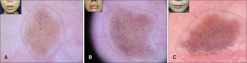
Fig. 1 (A~C) Pigmented cutaneous lesions located on lip in labial melanotic macules patients (inset). Dermoscopy revealed background brown pigmentation (indicated by red arrow), parallel lines (by purple arrows), circular lines (by blue arrows), and overlapping vessels (by orange arrows).
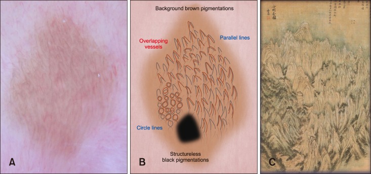
Fig. 2 (A~C) Schematic representations of dermoscopic patterns in patient with labial melanotic macules. (C) The ‘landscape painting pattern’ was derived from the 18th century Korean masterpiece, ‘General view of Mt. Geumgangsan’ by Jeong Seon. (B) The pattern was deemed to be present if the lesion satisfied all three of the following features; background brown pigmentation, parallel lines or circular lines, and overlapping vessels.
Reference
-
1. Ho KK, Dervan P, O'Loughlin S, Powell FC. Labial melanotic macule: a clinical, histopathologic, and ultrastructural study. J Am Acad Dermatol. 1993; 28:33–39. PMID: 8425968.
Article2. Gupta G, Williams RE, Mackie RM. The labial melanotic macule: a review of 79 cases. Br J Dermatol. 1997; 136:772–775. PMID: 9205516.
Article3. Mannone F, De Giorgi V, Cattaneo A, Massi D, De Magnis A, Carli P. Dermoscopic features of mucosal melanosis. Dermatol Surg. 2004; 30:1118–1123. PMID: 15274702.
Article4. Tsunemi Y, Saeki H, Tamaki K. Labial melanotic macule diagnosed by dermoscopy. Acta Derm Venereol. 2008; 88:524–525. PMID: 18779902.
Article5. Lin J, Koga H, Takata M, Saida T. Dermoscopy of pigmented lesions on mucocutaneous junction and mucous membrane. Br J Dermatol. 2009; 161:1255–1261. PMID: 19673880.
Article6. Blum A, Simionescu O, Argenziano G, Braun R, Cabo H, Eichhorn A, et al. Dermoscopy of pigmented lesions of the mucosa and the mucocutaneous junction: results of a multicenter study by the International Dermoscopy Society (IDS). Arch Dermatol. 2011; 147:1181–1187. PMID: 21680757.
- Full Text Links
-
- Actions
-
Cited
- CITED
-
- Close
- Share
-
- Similar articles
-
- Clinicopathologic Study of Labial Melanotic Macule
- Melanotic Schwannoma in Cervical Spine: A Case Report
- Dermoscopic Patterns of Onychomycosis: A Cross-sectional Study in One Institution
- Dermoscopic Findings and the Clinicopathologic Correlation of Pigmented Purpuric Dermatosis: A Retrospective Review of 60 Cases
- Labial Adhesions in Children: Report of Two Cases
