Is Anatomical Healing Essential for Better Clinical Outcome in Type II SLAP Repair? Clinico-Radiological Outcome after Type II SLAP Repair
- Affiliations
-
- 1Department of Orthopaedic Surgery, Seth GS Medical College and KEM Hospital, Mumbai, India.
- 2Department of Orthopedic Surgery, Seoul National University Bundang Hospital, Seoul National University College of Medicine, Seongnam, Korea. starsmstar@daum.net
- 3Department of Orthopaedic Surgery, St. George Hospital, Kogarah, Australia.
- KMID: 2418757
- DOI: http://doi.org/10.4055/cios.2018.10.3.358
Abstract
- BACKGROUND
We hypothesized that anatomical healing in superior labrum anterior to posterior (SLAP) repair is associated with good clinical outcome. The purposes of this study were to assess the failure rate of anatomical healing after arthroscopic repair of SLAP lesions using computed tomography arthrography (CTA), investigate correlation of the rate with clinical outcomes, and identify prognostic factors for anatomical failure following SLAP repair.
METHODS
We retrospectively evaluated the outcome of 43 patients at a minimum follow-up of 1 year after arthroscopic surgery for SLAP lesions or SLAP lesions associated with Bankart lesions. Twenty-eight patients underwent isolated SLAP repair and 15 patients underwent Bankart repair with SLAP repair. The anatomical outcome was assessed using CTA at 1 year after surgery. Clinical outcomes including visual analogue scale for pain and satisfaction and Constant score were assessed at the final follow-up. We investigated clinical failure that was defined as stiffness, loss of maximum rotation, deterioration of pain, and/or need for revision of surgery.
RESULTS
Anatomical failure occurred in 32.6% of patients (14/43), whereas 16.3% of patients (7/43) had clinical failure. Clinicoradiological assessment revealed that clinical failure occurred only in 7.1% of patients (1/14) with unhealed SLAP lesions, whereas it occurred in 20.7% of patients (6/29) with healed SLAP lesions. Isolated SLAP repair resulted in a higher risk of anatomical failure (risk ratio, 7.0) than combined SLAP repair (p = 0.015). Nonoverhead activities were associated with higher risk of anatomical failure (risk ratio, 2.9; p = 0.041). Patients above 35 years of age had more risk of anatomical failure (risk ratio, 3.5; p = 0.010). Clinical outcomes significantly improved regardless of anatomical failure (p < 0.001) and were not significantly different between unhealed and healed repairs (all p > 0.05).
CONCLUSIONS
Since patients with unhealed SLAP lesions had less clinical failure than patients with healed SLAP lesions, anatomical healing does not seem essential for better clinical outcome of SLAP II repair, especially in patients with higher healing failure risk (isolated SLAP repair, nonoverhead activities, and above 35 years of age). Therefore, we believe the indications of SLAP repair should be narrowed to avoid overtreatment.
Keyword
Figure
-
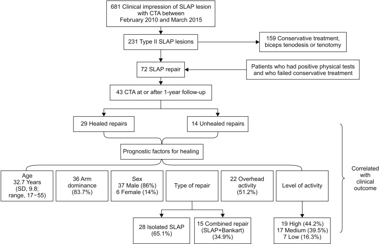
Fig. 1 Flow diagram of patient inclusion and data analysis. SLAP: superior labrum anterior to posterior, CTA: Computed tomography arthrography, SD: standard deviation.
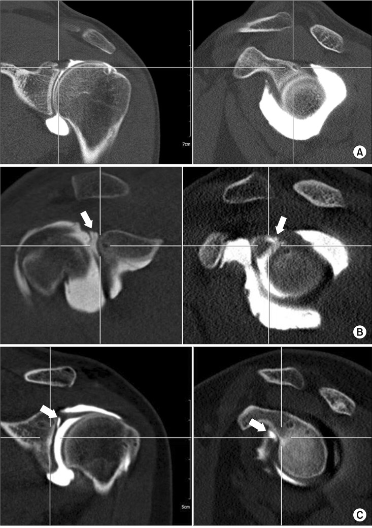
Fig. 2 Computed tomography arthrography (CTA) for superior labrum anterior to posterior (SLAP) repair evaluation. (A) An anatomically healed SLAP lesion after repair. (B) Anatomically unhealed SLAP lesion (arrows) after repair. Note the site of the dye leak between 10 and 12 o'clock positions. (C) CTA showing a healed lesion after repair that may be misinterpreted as an unhealed lesion (arrows). Note the site of leak beyond 12 o'clock.

Fig. 3 Superior labrum anterior to posterior (SLAP) repair. (A) Detection of a SLAP lesion (arrow). (B) SLAP repair.
Cited by 1 articles
-
National Trends in the Repair of Isolated Superior Labral Tear from Anterior to Posterior in Korea
Young-Hoon Jo, Hyun-Keun Oh, Soo-Young Jeong, Bong-Gun Lee
J Korean Med Sci. 2020;35(34):e285. doi: 10.3346/jkms.2020.35.e285.
Reference
-
1. Frank RM, Nho SJ, McGill KC, et al. Retrospective analysis of arthroscopic superior labrum anterior to posterior repair: prognostic factors associated with failure. Adv Orthop. 2013; 2013:125960. PMID: 23585969.
Article2. Brockmeyer M, Tompkins M, Kohn DM, Lorbach O. SLAP lesions: a treatment algorithm. Knee Surg Sports Traumatol Arthrosc. 2016; 24(2):447–455. PMID: 26818554.
Article3. Burkart A, Debski R, Musahl V, McMahon P, Woo SL. Biomechanical tests for type II SLAP lesions of the shoulder joint before and after arthroscopic repair. Orthopade. 2003; 32(7):600–607. PMID: 12883759.4. Schroder CP, Skare O, Reikeras O, Mowinckel P, Brox JI. Sham surgery versus labral repair or biceps tenodesis for type II SLAP lesions of the shoulder: a three-armed randomised clinical trial. Br J Sports Med. 2017; 51(24):1759–1766. PMID: 28495804.
Article5. Patterson BM, Creighton RA, Spang JT, Roberson JR, Kamath GV. Surgical trends in the treatment of superior labrum anterior and posterior lesions of the shoulder: analysis of data from the American Board of Orthopaedic Surgery Certification Examination Database. Am J Sports Med. 2014; 42(8):1904–1910. PMID: 24890780.6. Kim SH, Ha KI, Kim SH, Choi HJ. Results of arthroscopic treatment of superior labral lesions. J Bone Joint Surg Am. 2002; 84-A(6):981–985. PMID: 12063332.
Article7. Oh JH, Kim SH, Lee HK, Jo KH, Bae KJ. Trans-rotator cuff portal is safe for arthroscopic superior labral anterior and posterior lesion repair: clinical and radiological analysis of 58 SLAP lesions. Am J Sports Med. 2008; 36(10):1913–1921. PMID: 18495968.8. Oh JH, Lee HK, Kim JY, Kim SH, Gong HS. Clinical and radiologic outcomes of arthroscopic glenoid labrum repair with the BioKnotless suture anchor. Am J Sports Med. 2009; 37(12):2340–2348. PMID: 19776341.
Article9. Eime RM, Harvey JT, Charity MJ, Casey MM, Westerbeek H, Payne WR. Age profiles of sport participants. BMC Sports Sci Med Rehabil. 2016; 8:6. PMID: 26973792.
Article10. De Filippo M, Araoz PA, Pogliacomi F, et al. Recurrent superior labral anterior-to-posterior tears after surgery: detection and grading with CT arthrography. Radiology. 2009; 252(3):781–788. PMID: 19703862.
Article11. Kim SH, Choi JY, Yoo HJ, Hong SH. External rotation and active supination CT arthrography for the postoperative evaluation of type II superior labral anterior to posterior lesions. Knee Surg Sports Traumatol Arthrosc. 2016; 24(1):134–140. PMID: 25274092.
Article12. Oh JH, Kim JY, Choi JA, Kim WS. Effectiveness of multidetector computed tomography arthrography for the diagnosis of shoulder pathology: comparison with magnetic resonance imaging with arthroscopic correlation. J Shoulder Elbow Surg. 2010; 19(1):14–20. PMID: 19556150.
Article13. Choi BH, Kim NR, Moon SG, Park JY, Choi JW. Superior labral cleft after superior labral anterior-to-posterior tear repair: CT arthrographic features and correlation with clinical outcome. Radiology. 2016; 278(2):441–448. PMID: 26131912.
Article14. McCulloch PC, Andrews WJ, Alexander J, Brekke A, Duwani S, Noble P. The effect on external rotation of an anchor placed anterior to the biceps in type 2 SLAP repairs in a cadaveric throwing model. Arthroscopy. 2013; 29(1):18–24. PMID: 23177591.
Article15. Enad JG, Gaines RJ, White SM, Kurtz CA. Arthroscopic superior labrum anterior-posterior repair in military patients. J Shoulder Elbow Surg. 2007; 16(3):300–305. PMID: 17363292.
Article16. Kim DS, Yi CH, Yoon YS. Arthroscopic repair for combined Bankart and superior labral anterior posterior lesions: a comparative study between primary and recurrent anterior dislocation in the shoulder. Int Orthop. 2011; 35(8):1187–1195. PMID: 21369793.
Article17. Waterman BR, Arroyo W, Heida K, Burks R, Pallis M. SLAP repairs with combined procedures have lower failure rate than isolated repairs in a military population: surgical outcomes with minimum 2-year follow-up. Orthop J Sports Med. 2015; 3(8):2325967115599154. PMID: 26535389.18. Cho HL, Lee CK, Hwang TH, Suh KT, Park JW. Arthroscopic repair of combined Bankart and SLAP lesions: operative techniques and clinical results. Clin Orthop Surg. 2010; 2(1):39–46. PMID: 20191000.
Article19. Hantes ME, Venouziou AI, Liantsis AK, Dailiana ZH, Malizos KN. Arthroscopic repair for chronic anterior shoulder instability: a comparative study between patients with Bankart lesions and patients with combined Bankart and superior labral anterior posterior lesions. Am J Sports Med. 2009; 37(6):1093–1098. PMID: 19286910.20. Wilk KE, Macrina LC, Cain EL, Dugas JR, Andrews JR. The recognition and treatment of superior labral (slap) lesions in the overhead athlete. Int J Sports Phys Ther. 2013; 8(5):579–600. PMID: 24175139.21. Alpert JM, Wuerz TH, O'Donnell TF, Carroll KM, Brucker NN, Gill TJ. The effect of age on the outcomes of arthroscopic repair of type II superior labral anterior and posterior lesions. Am J Sports Med. 2010; 38(11):2299–2303. PMID: 20739578.
Article22. Mok D, Wang EL. Does age or gender of the patient influence the outcome of type II superior labrum anterior and posterior repair? Int J Shoulder Surg. 2012; 6(4):112–115. PMID: 23493640.
Article23. Cohen DB, Coleman S, Drakos MC, et al. Outcomes of isolated type II SLAP lesions treated with arthroscopic fixation using a bioabsorbable tack. Arthroscopy. 2006; 22(2):136–142. PMID: 16458798.
Article24. Alashkham A, Alraddadi A, Felts P, Soames R. Blood sup-ply and vascularity of the glenoid labrum: its clinical implications. J Orthop Surg (Hong Kong). 2017; 25(3):2309499017731632. PMID: 28920546.
Article25. Cook JL, Kiss ZS, Ptasznik R, Malliaras P. Is vascularity more evident after exercise? Implications for tendon imaging. AJR Am J Roentgenol. 2005; 185(5):1138–1140. PMID: 16247122.
Article
- Full Text Links
-
- Actions
-
Cited
- CITED
-
- Close
- Share
-
- Similar articles
-
- Arthroscopic Repair of Type II SLAP lesion with Bio-knotless Anchor
- New V-shaped Technique in SLAP Repair (Comparison of Cinical Results Between New V-shaped Repair and Conventional Rapair Technique in Arthroscopic Type II SLAP Surgery)
- Clinical Result of Arthroscopic Capsular Release and Repair for SLAP II Lesion with Stiffness
- Follow-up Result in Repairing a Type II Superior Labrum Anterior and Posterior (SLAP) Lesion when Associated with Shoulder Impingement Syndrome
- Arthroscopic Treatment of a Type II Superior Labrum Anterior to Posterior (SLAP) Lesion Combined with a Bankart Lesion: Comparative Study between Debridement and Repair of Type II SLAP Lesion by the Status of Lesion
