Comparison of treatment effects between four premolar extraction and total arch distalization using the modified C-palatal plate
- Affiliations
-
- 1Private Practice, Seoul, Korea.
- 2Department of Dentistry, College of Medicine, The Catholic University of Korea, Seoul, Korea.
- 3Department of Postgraduate Studies, the Universidad Autonóma del Paraguay, Asunción, Paraguay.
- 4School of Dentistry, University California San Francisco, San Francisco, CA, USA.
- 5Department of Orthodontics, Seoul St. Mary's Hospital, College of Medicine, The Catholic University of Korea, Seoul, Korea.
- 6Division of Orthodontics, Department of Dentistry, St. Vincent's Hospital, College of Medicine, The Catholic University of Korea, Seoul, Korea. seonghh@hotmail.com
- KMID: 2417964
- DOI: http://doi.org/10.4041/kjod.2018.48.4.224
Abstract
OBJECTIVE
The purpose of this study was to compare the skeletal, dental, and soft-tissue treatment effects of nonextraction therapy using the modified C-palatal plate (MCPP) to those of premolar extraction (PE) treatment in adult patients with Class II malocclusion.
METHODS
Pretreatment and posttreatment lateral cephalographs of 40 adult patients with Class II malocclusion were retrospectively analyzed. The MCPP group comprised 20 patients treated with total arch distalization of the maxillary arch while the PE group comprised 20 patients treated with four PE. Fifty-eight linear and angular measurements were analyzed to assess the changes before and after treatment. Descriptive statistics, paired t-test, and multivariate analysis of variance were performed to evaluate the treatment effects within and between the two groups.
RESULTS
The MCPP group presented 3.4 mm of retraction, 1.0 mm of extrusion, and 7.3° lingual inclination of the maxillary central incisor. In comparison, the PE group displayed greater amount of maxillary central incisor retraction and retroclination, mandibular incisor retraction, and upper lip retraction (5.3 mm, 14.8°, 5.1 mm, and 2.0 mm, respectively; p < 0.001 for all). In addition, the MCPP group showed 4.0 mm of distalization and 1.3 mm of intrusion with 2.9° distal tipping of the maxillary first molars.
CONCLUSIONS
These findings suggest the MCPP is an effective distalization appliance in the maxillary arch. The amount of incisor retraction, however, was significantly higher in the PE group. Therefore, four PE may be recommended when greater improvement of incisor position and soft-tissue profile is required.
Keyword
MeSH Terms
Figure
-

Figure 1 Linear cephalometric variables used for analyzing the effects of four premolar extraction or the modified C-palatal plate. Pt, Pterygoid; Or, orbitale; U, upper; L, lower; UL, upper lip; LL, lower lip; FH, Frankfort horizontal plane; HRL, horizontal reference line; VRL, vertical reference line; TVL, true vertical line; MeGo, menton to gonion line; Perp to MeGo, perpendicular line to MeGo passing the pogonion; 1, U1 crown to HRL; 2, U1 crown to VRL; 3, U1 apex to HRL; 4, U1 apex to VRL; 5, U6 crown to HRL; 6, U6 crown to VRL; 7, U6 apex to HRL; 8, U6 apex to VRL; 9, U7 crown to HRL; 10, U7 crown to VRL; 11, U7 apex to HRL; 12, U7 apex to VRL; 13, L1 crown to HRL; 14, L1 crown to VRL; 15, L1 apex to HRL; 16, L1 apex to VRL; 17, L6 crown to HRL; 18, L6 crown to VRL; 19, L6 apex to HRL; 20, L6 apex to VRL; 21, L7 crown to HRL; 22, L7 crown to VRL; 23, L7 apex to HRL; 24, L7 apex to VRL; 25, overjet; 26, overbite; 27, UL to TVL; 28, LL to TVL; 29, soft tissue A point to VRL; 30, soft tissue B point to VRL.
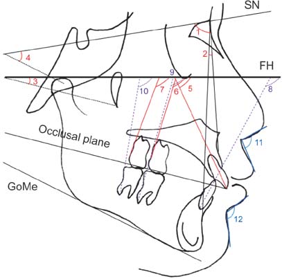
Figure 2 Angular cephalometric variables used for analyzing the effects of four premolar extraction or the modified C-palatal plate. SN, Sella-nasion; FH, Frankfort horizontal plane; Go, gonion; Me, menton; U, upper; L, lower; 1, SNA; 2, ANB; 3, occlusal plane angle; 4, SN to mandibular plane angle; 5, U1 to FH angle; 6, U6 to FH angle; 7, U7 to FH angle; 8, L1 to FH angle; 9, L6 to FH angle; 10, L7 to FH angle; 11, nasolabial angle; 12, mentolabial angle.

Figure 3 Linear and angular cephalometric variables in reference to the mandible for analyzing the effects of four premolar extraction or the modified C-palatal plate. Go, Gonion; Me, menton; mVRL, mandibular vertical reference line; Perp to, perpendicular to; L, lower; 1, L1 crown to mVRL; 2, L1 crown to GoMe; 3, L1 apex to mVRL; 4, L1 apex to GoMe; 5, L6 crown to mVRL; 6, L1 crown to GoMe; 7, L6 apex to mVRL; 8, L6 apex to GoMe; 9, L7 crown to mVRL; 10, L7 crown to GoMe; 11, L7 apex to mVRL; 12, L7 apex to GoMe; 13, L1 to GoMe angle; 14, L6 to GoMe angle; 15, L7 to GoMe angle.
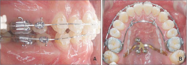
Figure 4 Mid-treatment oral photographs in the premolar extraction and nonextraction (modified C-palatal plate) groups. A, Extraction treatment; B, Nonextraction treatment.
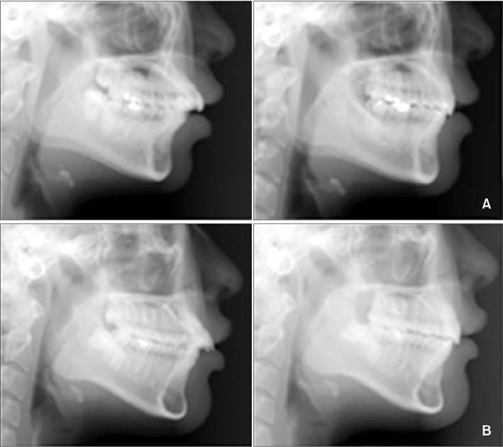
Figure 5 Representative changes in the lower third of the face based on the pretreatment (left) and posttreatment (right) lateral cephalographs in the premolar extraction and nonextraction groups. A, Extraction treatment; B, Nonextraction treatment.
Reference
-
1. Piao Y, Kim SJ, Yu HS, Cha JY, Baik HS. Five-year investigation of a large orthodontic patient population at a dental hospital in South Korea. Korean J Orthod. 2016; 46:137–145.
Article2. Fritz U, Diedrich P, Wiechmann D. Lingual technique--patients' characteristics, motivation and acceptance. Interpretation of a retrospective survey. J Orofac Orthop. 2002; 63:227–233.
Article3. Cardaropoli D, Gaveglio L, Abou-Arraj RV. Orthodontic movement and periodontal bone defects: Rationale, timing, and clinical implications. Semin Orthod. 2014; 20:177–187.
Article4. Peterson LJ, Ellis EE, Hupp JR, Tucker MR. Contemporary oral and maxillofacial surgery. 3rd ed. St. Louis, MO: Mosby;1998. p. 259–263.5. Crossman IG, Reed RT. Long term results of premolar extractions in orthodontic treatment. Br J Orthod. 1978; 5:61–66.
Article6. Garib DG, Bressane LB, Janson G, Gribel BF. Stability of extraction space closure. Am J Orthod Dentofacial Orthop. 2016; 149:24–30.
Article7. Cope JB. Temporary anchorage devices in orthodontics: A paradigm shift. Semin Orthod. 2005; 11:3–9.
Article8. Choi YJ, Lee JS, Cha JY, Park YC. Total distalization of the maxillary arch in a patient with skeletal Class II malocclusion. Am J Orthod Dentofacial Orthop. 2011; 139:823–833.
Article9. Kinzinger GS, Gülden N, Yildizhan F, Diedrich PR. Efficiency of a skeletonized distal jet appliance supported by miniscrew anchorage for noncompliance maxillary molar distalization. Am J Orthod Dentofacial Orthop. 2009; 136:578–586.
Article10. Bechtold TE, Kim JW, Choi TH, Park YC, Lee KJ. Distalization pattern of the maxillary arch depending on the number of orthodontic miniscrews. Angle Orthod. 2013; 83:266–273.
Article11. Sugawara J, Kanzaki R, Takahashi I, Nagasaka H, Nanda R. Distal movement of maxillary molars in nongrowing patients with the skeletal anchorage system. Am J Orthod Dentofacial Orthop. 2006; 129:723–733.
Article12. Mah SJ, Kim JE, Ahn EJ, Nam JH, Kim JY, Kang YG. Analysis of midpalatal miniscrew-assisted maxillary molar distalization patterns with simultaneous use of fixed appliances: A preliminary study. Korean J Orthod. 2016; 46:55–61.
Article13. Jung MH. A comparison of second premolar extraction and mini-implant total arch distalization with interproximal stripping. Angle Orthod. 2013; 83:680–685.
Article14. Sheridan JJ. Air-rotor stripping update. J Clin Orthod. 1987; 21:781–788.15. Sar C, Kaya B, Ozsoy O, Özcirpici AA. Comparison of two implant-supported molar distalization systems. Angle Orthod. 2013; 83:460–467.
Article16. Wilmes B, Nanda R, Nienkemper M, Ludwig B, Drescher D. Correction of upper-arch asymmetries using the Mesial-Distalslider. J Clin Orthod. 2013; 47:648–655.17. Papadopoulos MA. Orthodontic treatment of Class II malocclusion with miniscrew implants. Am J Orthod Dentofacial Orthop. 2008; 134:604.e1–604.e16.
Article18. Kircelli BH, Pektaş ZO, Kircelli C. Maxillary molar distalization with a bone-anchored pendulum appliance. Angle Orthod. 2006; 76:650–659.19. Escobar SA, Tellez PA, Moncada CA, Villegas CA, Latorre CM, Oberti G. Distalization of maxillary molars with the bone-supported pendulum: a clinical study. Am J Orthod Dentofacial Orthop. 2007; 131:545–549.
Article20. Kook YA, Kim SH, Chung KR. A modified palatal anchorage plate for simple and efficient distalization. J Clin Orthod. 2010; 44:719–730.21. Lee SK. Comparison of treatment effect between the MPAP and buccally miniscrew groups in nonextraction case [Master's thesis]. Seoul: The Catholic University of Korea;2016.22. Park CO, Sa'aed NL, Bayome M, Park JH, Kook YA, Park YS, et al. Comparison of treatment effects between the modified C-palatal plate and cervical pull headgear for total arch distalization in adults. Korean J Orthod. 2017; 47:375–383.
Article23. Grave K, Townsend G. Cervical vertebral maturation as a predictor of the adolescent growth spurt. Aust Orthod J. 2003; 19:25–32.24. Tweed CH. Indications for the extraction of teeth in orthodontic procedure. Am J Orthod Oral Surg. 1944–1945; 42:22–45.
Article25. Basciftci FA, Usumez S. Effects of extraction and nonextraction treatment on class I and class II subjects. Angle Orthod. 2003; 73:36–42.26. Saelens NA, De Smit AA. Therapeutic changes in extraction versus non-extraction orthodontic treatment. Eur J Orthod. 1998; 20:225–236.
Article27. Steyn CL, du Preez RJ, Harris AM. Differential premolar extractions. Am J Orthod Dentofacial Orthop. 1997; 112:480–486.
Article28. Duran GS, Görgülü S, Dindaroğlu F. Three-dimensional analysis of tooth movements after palatal miniscrew-supported molar distalization. Am J Orthod Dentofacial Orthop. 2016; 150:188–197.
Article29. Oh YH, Park HS, Kwon TG. Treatment effects of microimplant-aided sliding mechanics on distal retraction of posterior teeth. Am J Orthod Dentofacial Orthop. 2011; 139:470–481.
Article
- Full Text Links
-
- Actions
-
Cited
- CITED
-
- Close
- Share
-
- Similar articles
-
- Distalization with a modified C-palatal plate for severe upper crowding and a missing lower incisor
- Total arch distalization with interproximal stripping in a patient with severe crowding
- An experimental study of ideal arch form of the Class I malocclusion patients with first premolar extraction
- Comparison of treatment effects between the modified C-palatal plate and cervical pull headgear for total arch distalization in adults
- Osteometric Analysis of Palatal Bone Thickness for Orthodontic Miniscrew Placement
