Unilateral rostral mandibulectomy for gingival vascular hamartoma in two calves
- Affiliations
-
- 1Department of Veterinary Clinical Medicine, School of Veterinary Medicine, Tottori University, Tottori 680-8553, Japan. tsuka@muses.tottori-u.ac.jp
- 2Shimane Prefectural Federation Agricultural Mutual Aid Association, Shimane 690-0887, Japan.
- 3Tottori Prefectural Federation Agricultural Mutual Aid Association, Tottori 689-2202, Japan.
- KMID: 2417576
- DOI: http://doi.org/10.4142/jvs.2018.19.4.582
Abstract
- A 2-month-old female Holstein calf and a 5-month-old female Japanese black calf presented with gingival vascular hamartoma located in the interdental space between the second and third mandibular incisors in the right and left mandibles, respectively. On radiographic or computed tomographic images, osteolytic changes appeared within the mandibular bones adjacent to the masses. The masses were removed along with affected mandibular bone by using unilateral rostral mandibulectomy. After surgery, both cases exhibited a normal appetite and grew normally, with no cosmetic changes or recurrences. Unilateral rostral mandibulectomy can be applied for invasive gingival vascular hamartomas associated with osteolytic changes.
MeSH Terms
Figure
-

Fig. 1 Gross appearance (A) and computed tomographic (CT) image (B) of the right rostral aspect of the mandible, and postoperative CT image of the mandible (C) in Case 1. (B) The enlarged mass (*) overlaying the second (I2) and third incisors (I3) resulted in dislocation of the third incisor laterally within the right mandible. Osteolysis is evident within the rostral mandibular bone surrounding the roots of the incisor and canine. (C) After surgery, intact removal of the affected right mandible together with the mass and four teeth appears due to the oblique and straight cutting. I1, first incisor; I2, second incisor; I3, third incisor; C, canine teeth. Scale bar = 5 mm on CT images.
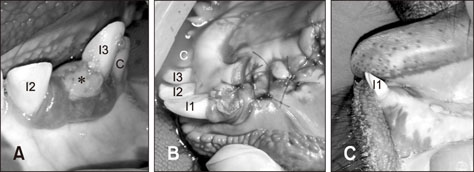
Fig. 2 Gross appearances of the left rostral aspect of the mandible at admission (A), soon after surgery (B), and at 6 months after surgery (C) in Case 2. A 2-cm diameter, dark-reddish mass (*) appears within the interdental space between the second (I2) and third incisors (I3), and there is a well-demarcated margin between mass and gingiva. The rostral and sublingual gingival mucosa are apposed over the cut surface of the bone with absorbable suture material. The surgical wound is intact and closed without recurrence (C). I1, first incisor; I2, second incisor; I3, third incisor; C, canine teeth.
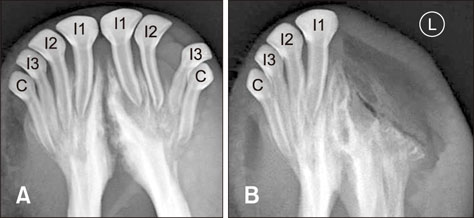
Fig. 3 Ventrodorsal intraoral radiographs of the mandible taken at admission (A) and the day after surgery (B) in Case 2. The interdental space between the second and third incisors was wider. Osteolysis was evident within the roots of the incisor and canine tooth. Most of the osteolytic region of the mandible bone was removed. I1, first incisor; I2, second incisor; I3, third incisor; C, canine teeth.
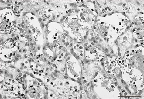
Fig. 4 Histopathological photomicrograph of the mass removed from Case 2. Hypertrophied endothelial cells have formed vascular lumens containing erythrocytes and fibrinous material. The interstitial tissue was edematous, with many eosinophils. H&E stain. 400×. Scale bar = 50 µm.
Reference
-
1. Aspinall V, Cappello M. Comparative anatomy and physiology. In : Aspinall V, Cappello M, editors. Introduction to Veterinary Anatomy and Physiology Textbook. 3rd ed. London: Elsevier Health Sciences;2015. p. 211–244.2. Mendez-Angulo JL, Tatarniuk DM, Ruiz I, Ernst N. Extensive rostral mandibulectomy for treatment of ameloblastoma in a horse. Vet Surg. 2014; 43:222–226.
Article3. Mohammadi GR, Maleki M, Sardari K. Gingival vascular hamartoma in a young Holstein calf. Comp Clin Pathol. 2007; 16:73–75.
Article4. Rösti L, Lauper J, Merhof K, Gorgas D, Ross S, Grest P, Welle MM. [Blood vessel anomalities in the oral cavity of two calves]. Schweiz Arch Tierheilkd. 2013; 155:627–632. German.5. Sheahan BJ, Donnelly WJ. Vascular hamartomas in the gingiva of two calves. Vet Pathol. 1981; 18:562–564.
Article6. Taney KG, Dubielzig RR, Trotter TS, Smith MM. Bilateral maxillary periodontal ligament hamartoma in a dog. J Vet Dent. 2005; 22:91–95.
Article7. Tetens J, Ross MW, Sweeney RW. Rostral mandibulectomy for treatment of an ameloblastic fibro-odontoma in a cow. J Am Vet Med Assoc. 1995; 207:1616–1617.8. Verstraete FJ. Mandibulectomy and maxillectomy. Vet Clin North Am Small Anim Pract. 2005; 35:1009–1039. viii
Article9. Wilson RB. Gingival vascular hamartoma in three calves. J Vet Diagn Invest. 1990; 2:338–339.
Article10. Yeruham I, Abramovitch I, Perl S. Gingival vascular hamartoma in two calves. Aust Vet J. 2004; 82:152–153.
Article
