Mucinous Adenocarcinoma Arising within a Colonic Diverticulum Mimicking a Diverticular Abscess: A Case Report
- Affiliations
-
- 1Department of Radiology, Soonchunhyang University College of Medicine, Seoul Hospital, Seoul, Korea. jy0707hwang@schmc.ac.kr
- 2Department of Surgery, Soonchunhyang University College of Medicine, Seoul Hospital, Seoul, Korea.
- 3Department of Pathology, Soonchunhyang University College of Medicine, Seoul Hospital, Seoul, Korea.
- KMID: 2416404
- DOI: http://doi.org/10.3348/jksr.2018.79.1.40
Abstract
- Colon cancer arising in a colonic diverticulum is very rare. There are only a few reported cases of colon cancer associated with a diverticulum. Of these reported cases, only a few are those of a mucinous adenocarcinoma. Here, we report a case of an 82-year-old female with a mucinous adenocarcinoma arising in the ascending colonic diverticulum, which clinically and radiologically mimicked perforated diverticulitis with abscess formation. Although such cases are rare, our findings suggest that malignant tumors may be misdiagnosed as diverticular diseases and should be considered during work-up.
MeSH Terms
Figure
-
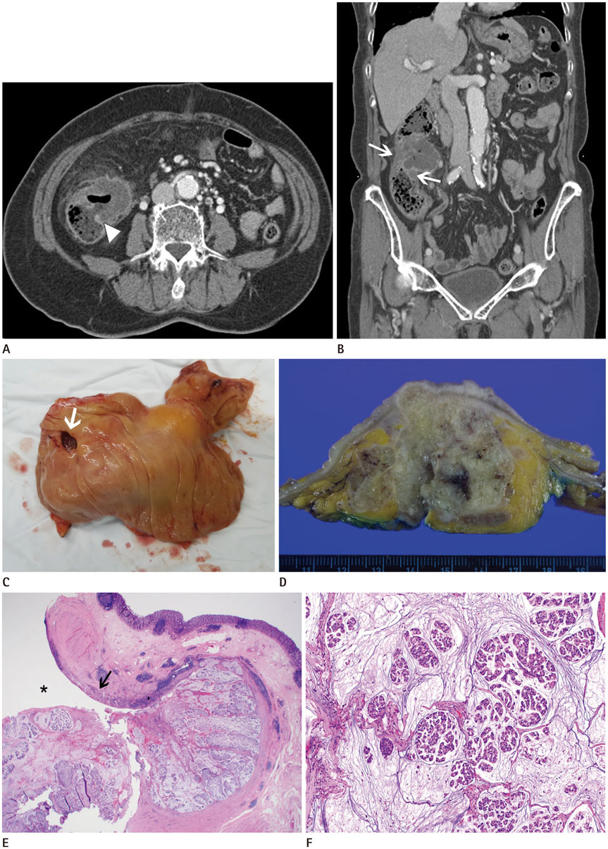
Fig. 1 A 82-year-old female with a mucinous adenocarcinoma arising in ascending colonic diverticulum. A. Axial contrast-enhanced abdominal CT showing a peripheral-enhancing, low-attenuated, exophytic mass at the medial wall of the proximal ascending colon. The mass is connected to the lumen of the proximal ascending colon via an out-pouching sac (arrowhead), indicating a colonic diverticulum. Mild soft tissue stranding was noted in the surrounding fat. B. In the coronal reformatted image, the involved segment of the colon shows mural thickening with preservation of the layering pattern (arrows). The appendix is unremarkable. C. The opened lumen of the colon specimen showing a large diverticular opening (arrow) with intact overlying mucosa. D. Cut section revealed an irregular mass in pericolic soft tissue, involving the colonic wall (6.0 × 5.5 cm). E. The mucosa lining the diverticulum is eroded (arrow) along the diverticular opening (asterisk). An abundant mucin pool with tumor cell nests was present in the deeper portion of the diverticulum (hematoxylin and eosin stain, × 12.5). F. The mass has floating, single, or nested tumor cells in the mucin pool (hematoxylin and eosin stain, × 100).
Reference
-
1. Jacobs DO. Clinical practice. diverticulitis. N Engl J Med. 2007; 357:2057–2206.2. Tolley JA 3rd. Chronic diverticulitis with perforation and associated carcinoma of the cecum. Dis Colon Rectum. 1967; 10:389–393.
Article3. Kwon JH, Han KH, Chang WS, Nam KH, Han MS, Ahn JH, et al. A case of a mucinous adenocarcinoma arising from a rectal diverticulum. J Korean Soc Coloproctol. 2012; 28:222–224.
Article4. Kikuchi T, Kotanagi H, Kon H, Koyama K, Ito S, Otaka M. Mucosal carcinoma within a colonic diverticulum. J Gastroenterol. 1999; 34:622–625.
Article5. Soliman ML, Cerda SR, Xu H. Invasive moderately-differentiated mucinous adenocarcinoma incidentally identified in perforated acute diverticulitis with abscess formation. Pathol Lab Med Open J. 2016; 1:32–36.6. Mayo WJ. Diverticulitis of the large intestine. JAMA. 1917; 781–785.
Article7. Rowe RJ, Kollmar GH. Diverticulitis of the colon complicated by carcinoma. Int Abstr Surg. 1952; 94:1–9.8. Park JS, Huh JW, Park YA, Cho YB, Yun SH, Kim HC, et al. Prognostic comparison between mucinous and nonmucinous adenocarcinoma in colorectal cancer. Medicine (Baltimore). 2015; 94:e658.
Article9. Hussain SM, Outwater EK, Siegelman ES. Mucinous versus nonmucinous rectal carcinomas: differentiation with MR imaging. Radiology. 1999; 213:79–85.
Article10. Chintapalli KN, Chopra S, Ghiatas AA, Esola CC, Fields SF, Dodd GD 3rd. Diveriticulitis versus colon cancer: differentiation with helical CT findings. Radiology. 1999; 210:429–435.
Article
- Full Text Links
-
- Actions
-
Cited
- CITED
-
- Close
- Share
-
- Similar articles
-
- A Case of a Mucinous Adenocarcinoma Arising from a Rectal Diverticulum
- A Case of Mucinous Adenocarcinoma of the Colon Presenting with Psoas Abscess
- Mucinous Adenocarcinoma Arising at the Anastomotic Site after Operation for Hirschsprung's Disease: Case Report
- Clinical Characteristics of Colonic Diverticular Disease Diagnosed with Colonoscopy
- Mucinous Adenocarcinoma Arising in the Renal Pelvis: A Case Report
