Adenosquamous Carcinoma of Duodenal Bulb Mimicking Subepithelial Tumor on Computed Tomography: A Case Report
- Affiliations
-
- 1Department of Radiology, Kangnam Sacred Heart Hospital, Hallym University College of Medicine, Seoul, Korea. baccas@hallym.or.kr
- 2Department of Pathology, Kangnam Sacred Heart Hospital, Hallym University College of Medicine, Seoul, Korea.
- 3Department of General Surgery, Kangnam Sacred Heart Hospital, Hallym University College of Medicine, Seoul, Korea.
- KMID: 2416394
- DOI: http://doi.org/10.3348/jksr.2018.79.2.92
Abstract
- Adenosquamous carcinomas of the duodenum are extremely rare neoplasms in which both glandular and squamous elements demonstrate malignant characteristics. Few cases of adenosquamous carcinoma involving the second or third segment of the duodenum have been reported in the literature. Herein, we report the first case of adenosquamous carcinoma of the bulb of the duodenum that mimicked subepithelial tumor on computed tomography in a 59-year-old man.
Figure
-
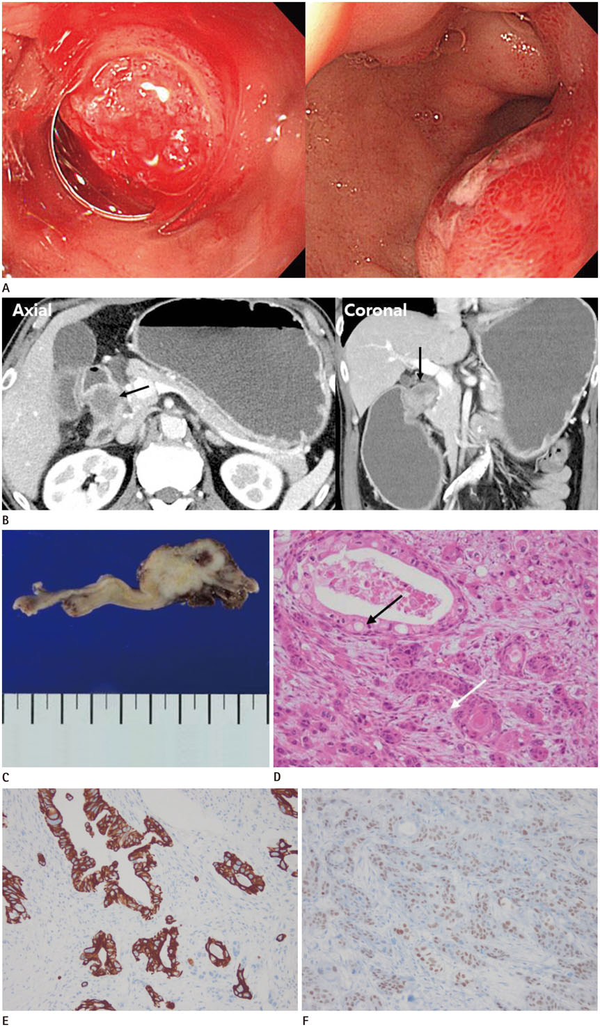
Fig. 1 A 59-year-old man with adenosquamous carcinoma involving duodenal bulb. A. Upper gastrointestinal endoscopy reveals a subepithelial mass at duodenal bulb and spurting bleeding on the surface of the mass (left), and epinephrine injection is done for hemostasis (right). B. Axial and coronal images of contrast enhanced CT scan show a 3 cm sized well-circumscribed hypoattenuating mass surrounded by hyperattenuating duodenal wall at the bulb of duodenum (arrows). The lesion causes gastric outlet obstruction. C. Grossly, a well-defined solid mass shows granular, necrotic, and hemorrhagic cut surface involves the duodenal wall. D. Microscopically, the lesion displays glands with intracellular mucin (black arrow) and islands with intercellular bridges and keratin pearl typical of squamous differentiation (white arrow) (hematoxylin and eosin stain, × 100). E. Immunohistochemically, the glandular part is immunoreactive for cytokeratin 7 (× 200). F. The squamous part is positive for p40 (× 200).
Reference
-
1. Daga G, Kerkar P. Adenosquamous carcinoma of the duodenum: a rare entity. Indian J Surg Oncol. 2016; 7:470–474.
Article2. Hammami MB, Chhaparia A, Piao J, Zhou Y, Hachem C, Lai J. Mixed adenocarcinoma and squamous cell carcinoma of duodenum: a case report and review of the literature. Case Rep Gastroenterol. 2017; 11:402–410.
Article3. de la Cruz A, de la Cruz E, Sanchez MJ, Ortiz S, Lobato A, Merino E. Adenosquamous carcinoma of the duodenum. an immunohistochemical study. Pathol Res Pract. 1993; 189:481–485. discussion 485-487.4. Takayoshi K, Ariyama H, Tamura S, Yoda S, Arita T, Yamaguchi T, et al. Intraluminal superior vena cava metastasis from adenosquamous carcinoma of the duodenum: a case report. Oncol Lett. 2016; 11:605–609.
Article5. Yang SJ, Ooyang CH, Wang SY, Liu YY, Kuo IM, Liao CH, et al. Adenosquamous carcinoma of the ampulla of Vater - a rare disease at unusual location. World J Surg Oncol. 2013; 11:124.
Article6. Ueno N, Sano T, Kanamaru T, Tanaka K, Nishihara T, Idei Y, et al. Adenosquamous cell carcinoma arising from the papilla major. Oncol Rep. 2002; 9:317–320.
Article7. Kshirsagar AY, Nangare NR, Vekariya MA, Gupta V, Pednekar AS, Wader JV, et al. Primary adenosquamous carcinoma of ampulla of Vater-a rare case report. Int J Surg Case Rep. 2014; 5:393–395.
Article8. Hoshimoto S, Aiura K, Shito M, Kakefuda T, Sugiura H. Adenosquamous carcinoma of the ampulla of Vater: a case report and literature review. World J Surg Oncol. 2015; 13:287.
Article9. Hatzaras I, Palesty JA, Abir F, Sullivan P, Kozol RA, Dudrick SJ, et al. Small-bowel tumors: epidemiologic and clinical characteristics of 1260 cases from the connecticut tumor registry. Arch Surg. 2007; 142:229–235.10. Goldner B, Stabile BE. Duodenal adenocarcinoma: why the extreme rarity of duodenal bulb primary tumors? Am Surg. 2014; 80:956–959.
Article
- Full Text Links
-
- Actions
-
Cited
- CITED
-
- Close
- Share
-
- Similar articles
-
- A Case of Tubular Adenocarcinoma on Fistula of Duodenal Bulb
- A Case of Primary Aortoenteric Fistula Mimicking Duodenal Subepithelial Tumor
- A Case of Adenosquamous Carcinoma of the Ampulla of Vater
- Adenosquamous Carcinoma of the Ascending Colon: A Case Report and Review of the Literature
- A Case of Cutaneous Metastatic Adenosquamous Carcinoma of the Pancreas
