Obstet Gynecol Sci.
2018 May;61(3):395-403. 10.5468/ogs.2018.61.3.395.
Doppler sonography of perifibroid and intrafibroid arteries of uterine leiomyomas
- Affiliations
-
- 1Department of Radiology, Obafemi Awolowo University Teaching Hospitals Complex, Ile-Ife, Nigeria. ibmcontacts@gmail.com
- KMID: 2416129
- DOI: http://doi.org/10.5468/ogs.2018.61.3.395
Abstract
OBJECTIVE
To sonographically evaluate the dominant fibroid nodule vascularity and flow velocity pattern of perifibroid and intrafibroid arteries.
METHODS
We recruited 140 women with uterine fibroids. Their uteri were scanned to determine the vascularity of fibroid nodules and the Doppler indices of the fibroid arteries.
RESULTS
The median volume of the dominant leiomyoma nodule was 133 cm3 (range=1.5-2,575 cm3). Eighty-three subjects (59.3%) had a dominant leiomyoma nodule volume of ≤200.0 cm3 while the volume of the dominant leiomyoma nodule was >200.0 cm3 in 57 (40.7%) subjects. The dominant fibroid nodule was vascular in 137 (97.9%) subjects and avascular in 3 (2.1%). All the perifibroid artery indices (except the end-diastolic velocity [EDV] and diastolic average ratio [DAR]) are significantly higher than those of the intrafibroid artery. The mean Doppler indices of perifibroid vs. intrafibroid arteries as follows: peak systolic velocity (PSV; 52.1 vs. 45.4 cm/s); EDV (21.1 vs. 22.4 cm/s); time-averaged maximum velocity (TAMX; 31.5 vs. 30.4 cm/s); time- averaged mean velocity (Tmean; 14.3 vs. 13.8 cm/s); pulsatility index (PI; 1.1 vs. 0.8); resistive index (RI; 0.6 vs. 0.5); systolic-diastolic ratio (SDR; 2.7 vs. 2.1); impedance index (ImI; 2.7 vs. 2.1); and DAR (0.66 vs. 0.74); P < 0.001 for all indices.
CONCLUSION
The predominant pattern of fibroid vascularity is peripheral vascularity and the perifibroid artery indices (except EDV and DAR) are significantly higher than those of the intrafibroid artery. Recurrent fibroids in women with previous myomectomy had significantly higher intrafibroid PI, RI, SDR, and ImI than those without previous myomectomy.
MeSH Terms
Figure
-
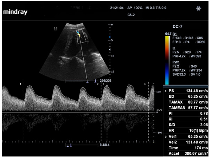
Fig. 1 Longitudinal triplex sonogram of a myomatous uterus showing the waveform pattern and Doppler indices of a perifibroid (peripheral) artery.
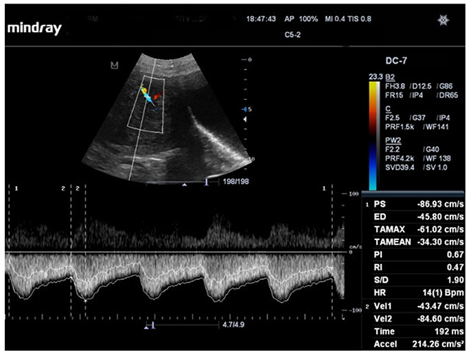
Fig. 2 Longitudinal triplex sonogram of a myomatous uterus showing the waveform pattern and Doppler indices of an intrafibroid (core) artery.
Reference
-
1. Olivetti L, Grazioli L, Frittoli B. Diagnostic imaging of the uterus. In : Olivetti L, Grazioli L, editors. Imaging of urogenital diseases: a color atlas. Milan: Springer-Verlag Italia;2009. p. 367–409.2. Kjerulff KH, Langenberg P, Seidman JD, Stolley PD, Guzinski GM. Uterine leiomyomas: racial differences in severity, symptoms and age at diagnosis. J Reprod Med. 1996; 41:483–490.3. Cramer SF, Patel A. The frequency of uterine leiomyomas. Am J Clin Pathol. 1990; 94:435–438.
Article4. Stewart EA, Nowak RA. Leiomyoma-related bleeding: a classic hypothesis updated for the molecular era. Hum Reprod Update. 1996; 2:295–306.
Article5. Sampson JA. The blood supply of uterine myomata. Surg Gynecol Obstet. 1912; 14:215–230.6. Farrer-Brown G, Beilby JO, Tarbit MH. Venous changes in the endometrium of leiomyomatous uteri. Obstet Gynecol. 1971; 38:743–751.7. Farrer-Brown G, Beilby JO, Rowles PM. Microvasculature of the uterus: an injection method of study. Obstet Gynecol. 1970; 35:21–30.8. Aitken E, Khaund A, Hamid SA, Millan D, Campbell S. The normal human myometrium has a vascular spatial gradient absent in small leiomyomas. Hum Reprod. 2006; 21:2669–2678.9. Fleischer AC. Color Doppler sonography of uterine disorders. Ultrasound Q. 2003; 19:179–189.
Article10. Weintraub JL, Romano WJ, Kirsch MJ, Sampaleanu DM, Madrazo BL. Uterine artery embolization: sonographic imaging findings. J Ultrasound Med. 2002; 21:633–637.11. Bhatt S, Dogra V. Doppler imaging of the uterus and adnexae. Ultrasound Clin. 2006; 1:201–221.
Article12. Sohaey R, Larsen SA. Uterine leiomyoma. In : Ahuja AT, Griffith JF, Wong KT, Antonio GE, Chu WC, editors. Diagnostic imaging: ultrasound. 1st ed. Salt Lake City (UT): Amrisys Inc.;2007.13. Chang T. Uterine leiomyomas [Internet]. c2010. cited 2013 Mar 5. Available from: http://www.drtchang.com.au.14. Smith B. Modern trends in gynecological ultrasound: a clinical and technical review. Ultrasound. 2006; 14:6–19.15. Tsuda H, Kawabata M, Nakamoto O, Yamamoto K. Clinical predictors in the natural history of uterine leiomyoma: preliminary study. J Ultrasound Med. 1998; 17:17–20.
Article16. Myers SL, Baird DD, Olshan AF, Herring AH, Schroeder JC, Nylander-French LA, et al. Self-report versus ultrasound measurement of uterine fibroid status. J Womens Health (Larchmt). 2012; 21:285–293.
Article17. Zivković N, Zivković K, Despot A, Paić J, Zelić A. Measuring the volume of uterine fibroids using 2- and 3-dimensional ultrasound and comparison with histopathology. Acta Clin Croat. 2012; 51:579–589.18. Alataş G, Aksoy E, Akarsu C, Yakin K, Bahçeci M. The effect of uterine volume on uterine artery Doppler velocimetry in the myomatous state. Gynecol Obstet Invest. 1997; 43:55–59.
Article19. Vetter K, Gonser M, Voigt HJ. Indices for the evaluation of Doppler sonograms. In : Sohn C, Voigt HJ, Vetter K, editors. Doppler ultrasound in gynecology and obstetrics. Stuttgart: Georg Thieme Verlag;2004. p. 29–39.20. Maulik D, Yarlagadda P, Youngblood JP, Ciston P. Comparative efficacy of umbilical arterial Doppler indices for predicting adverse perinatal outcome. Am J Obstet Gynecol. 1991; 164:1434–1440.
Article21. Pelage JP, Cazejust J, Pluot E, Le Dref O, Laurent A, Spies JB, et al. Uterine fibroid vascularization and clinical relevance to uterine fibroid embolization. Radiographics. 2005; 25:Suppl 1. S99–117.
Article22. Sosić A, Skupski DW, Streltzoff J, Yun H, Chervenak FA. Vascularity of uterine myomas: assessment by color and pulsed Doppler ultrasound. Int J Gynaecol Obstet. 1996; 54:245–250.23. Szabó I, Szánthó A, Csabay L, Csapó Z, Szirmai K, Papp Z. Color Doppler ultrasonography in the differentiation of uterine sarcomas from uterine leiomyomas. Eur J Gynaecol Oncol. 2002; 23:29–34.24. Kurjak A, Kupesic-Urek S, Miric D. The assessment of benign uterine tumor vascularization by transvaginal color Doppler. Ultrasound Med Biol. 1992; 18:645–649.
Article25. Samani FG, Jabbary R, Mashrabi O. Study of uterine artery blood flow in leiomyomatous uterus. Life Sci J. 2012; 9:583–586.26. Idowu BM, Ibitoye BO, Adetiloye VA. Uterine artery Doppler velocimetry of uterine leiomyomas in Nigerian women. Rev Bras Ginecol Obstet. 2017; 39:464–470.
Article27. Weston G, Healy DL. Uterine fibroid pseudocapsule [Internet]. [palce unknown]: Obgynkey;2016. Sep. 20. cited 2017 May 6. Available from: http://obgynkey.com/uterine-fibroid-pseudocapsule/.28. McLucas B, Perrella R, Goodwin S, Adler L, Dalrymple J. Role of uterine artery Doppler flow in fibroid embolization. J Ultrasound Med. 2002; 21:113–120.
Article29. Tranquart F, Brunereau L, Cottier JP, Marret H, Gallas S, Lebrun JL, et al. Prospective sonographic assessment of uterine artery embolization for the treatment of fibroids. Ultrasound Obstet Gynecol. 2002; 19:81–87.
Article30. Fleischer AC, Donnelly EF, Campbell MG, Mazer MJ, Grippo D, Lipsitz NL. Three-dimensional color Doppler sonography before and after fibroid embolization. J Ultrasound Med. 2000; 19:701–705.
Article31. Sladkevicius P, Valentin L, Marsal K. Transvaginal Doppler examination of uteri with myomas. J Clin Ultrasound. 1996; 24:135–140.
Article32. Tsiligiannis SE, Zaitseva M, Coombs PR, Shekleton P, Olshansky M, Hickey M, et al. Fibroid-associated heavy menstrual bleeding: correlation between clinical features, Doppler ultrasound assessment of vasculature, and tissue gene expression profiles. Reprod Sci. 2013; 20:361–370.
Article33. Chiang CH, Chang MY, Hsu JJ, Chiu TH, Lee KF, Hsieh TT, et al. Tumor vascular pattern and blood flow impedance in the differential diagnosis of leiomyoma and adenomyosis by color Doppler sonography. J Assist Reprod Genet. 1999; 16:268–275.34. Creighton S, Bourne TH, Lawton FG, Crayford TJ, Vyas S, Campbell S, et al. Use of transvaginal ultrasonography with color Doppler imaging to determine an appropriate treatment regimen for uterine fibroids with a GnRH agonist before surgery: a preliminary study. Ultrasound Obstet Gynecol. 1994; 4:494–498.
Article35. Yosry LM, Hamed E, El Naggar AA. The effect of aromatase inhibitor on uterine leiomyoma volume by ultrasonography and color Doppler. J Am Sci. 2012; 8:1031–1034.36. Huang SC, Yu CH, Huang RT, Hsu KF, Tsai YC, Chou CY. Intratumoral blood flow in uterine myoma correlated with a lower tumor size and volume, but not correlated with cell proliferation or angiogenesis. Obstet Gynecol. 1996; 87:1019–1024.37. Testa AC, Pomini F, Fattorossi A, Battaglia A, Ferrandina G, Mansueto D, et al. Doppler velocimetry and cytofluorimetric analysis in uterine myomas. Gynecol Obstet Invest. 2003; 56:139–142.
- Full Text Links
-
- Actions
-
Cited
- CITED
-
- Close
- Share
-
- Similar articles
-
- Role of Color Doppler US in the Evaluation of Uterine Leiomyoma Treated with Gonadotrophin-Releasing Hormone (GnRH) Agonist (Zoladex)
- Are Transvaginal Sonography and Color doppler findings Useful for the Diagnosis of Adenomyosis?
- Abnormal Uterine Bleeding Due to Vascular Abnormality Caused by D&E : Doppler Sonography for Diagnosis and Transcatheter Arterial Embolization for Treatment
- Role of Intraoperative Microvascular Doppler Sonography in the Surgery of Cerebral Aneurysm
- Transvaginal Doppler Ultrasonography of Uterine Intraendometrial and Intramyometrial Arteries as a Predictor of Pregnancy after In Vitro Fertilization and Embryo Transfer
