Clinical Application of 2017 McDonald Diagnostic Criteria for Multiple Sclerosis
- Affiliations
-
- 1Neurological Unit, MS Centre, A. Manzoni Hospital-ASST Lecco, Lecco, Italy. vittorio.mantero@hotmail.com
- 2Neurological Unit, ASST Valtellina Alto Lario, Sondrio, Italy.
- 3Unit of Neurorehabilitation, Multiple Sclerosis Center, I.R.C.C.S. Santa Maria Nascente-Fondazione Don Gnocchi, Milano, Italy.
- KMID: 2415057
- DOI: http://doi.org/10.3988/jcn.2018.14.3.387
Abstract
- BACKGROUND AND PURPOSE
McDonald criteria for multiple sclerosis diagnosis have been revised over the years, diagnostic procedures have been simplified and earlier diagnosis facilitated. The new 2017 revision introduces other important changes, with a further simplification for the diagnosis. Oligoclonal bands reassume a more relevant role in the workup.
METHODS
We describe 3 typical cases of patients admitted for clinically isolated syndrome and illustrate how the application of the new criteria can change the diagnostic approach with respect to the previous criteria.
RESULTS
In two of the three cases a diagnosis of multiple sclerosis is now possible.
CONCLUSIONS
The new 2017 Multiple Sclerosis criteria may have an important impact in clinical practice with an earlier treatment to avoid the risk of disease dissemination. Their application requires a careful assessment to avoid misdiagnosis and mistreatments.
Keyword
Figure
-
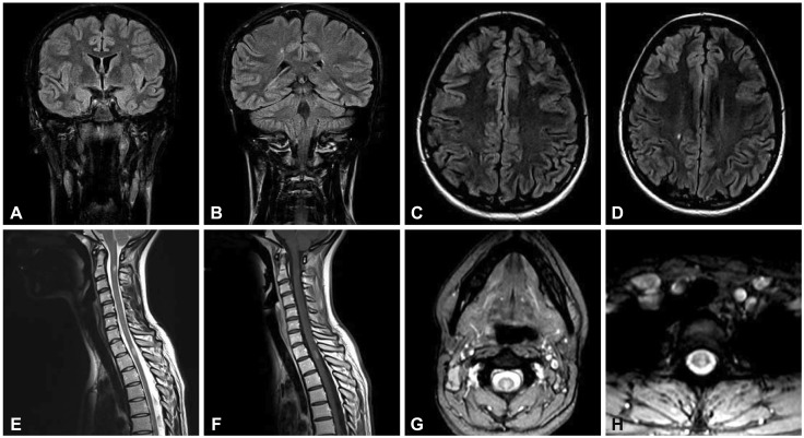
Fig. 1 Brain coronal plane FLAIR-weighted and axial plane T2-weighted scan showing hyperintense lesions in the left frontal lobe (A and C) and in right centrum semiovale (B and D), spinal cord sagittal and axial plane T2-weighted showing hyperintense lesions at C2 (E and G), and D1–D2 level (E and H), with enhancement on T1 post-Gadolinium scan (F).
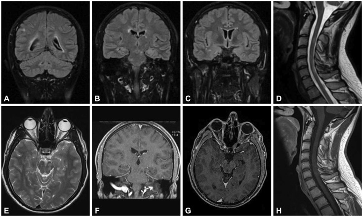
Fig. 2 Brain coronal plane FLAIR-weighted scan showing hyperintense juxtacortical (A), periventricular and infratentorial lesions (B and C) without enhancement on T1 post-Gadolinium scan (F and G), axial plane T2-weighted scan showing slight signal hyperintensity in left optic nerve (E), spinal cord sagittal plane T2-weighted showing hyperintense lesions at multiple levels (D) without enhancement on T1 post-Gadolinium scan (H).
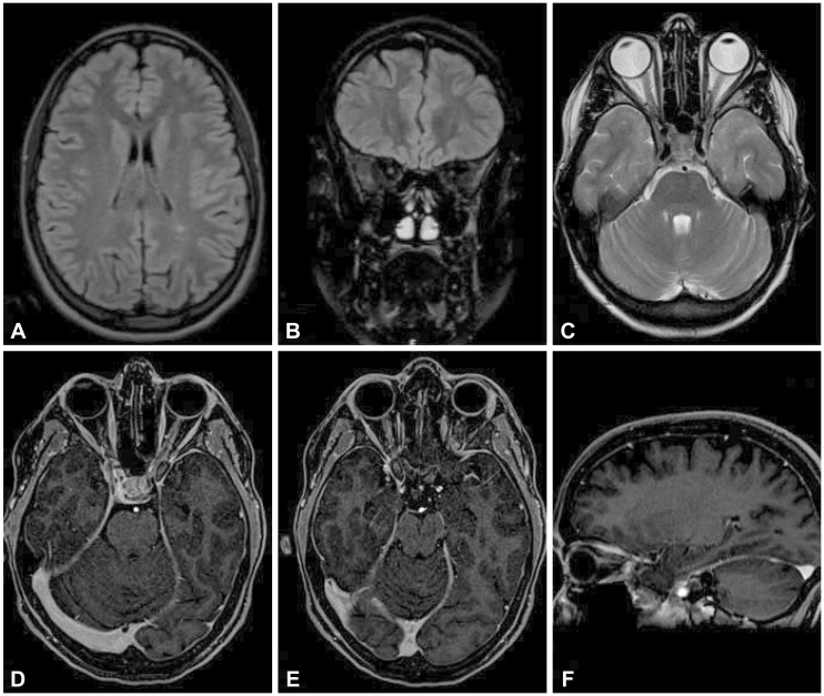
Fig. 3 Brain axial plane FLAIR-weighted scan showing hyperintensity of the left peritrigonal white matter (A) and coronal and axial plane FLAIR-weighted scan showing slight hyperintensity in the right optic nerve (B and C) without clear enhancement on T1 post-Gadolinium axial and sagittal scan (D, E, and F).
Reference
-
1. Miller DH, Chard DT, Ciccarelli O. Clinically isolated syndromes. Lancet Neurol. 2012; 11:157–169. PMID: 22265211.
Article2. Brownlee WJ, Miller DH. Clinically isolated syndromes and the relationship to multiple sclerosis. J Clin Neurosci. 2014; 21:2065–2071. PMID: 25027666.
Article3. McDonald WI, Compston A, Edan G, Goodkin D, Hartung HP, Lublin FD, et al. Recommended diagnostic criteria for multiple sclerosis: guidelines from the International Panel on the diagnosis of multiple sclerosis. Ann Neurol. 2001; 50:121–127. PMID: 11456302.
Article4. Polman CH, Reingold SC, Edan G, Filippi M, Hartung HP, Kappos L, et al. Diagnostic criteria for multiple sclerosis: 2005 revisions to the “McDonald criteria”. Ann Neurol. 2005; 58:840–846. PMID: 16283615.
Article5. Polman CH, Reingold SC, Banwell B, Clanet M, Cohen JA, Filippi M, et al. Diagnostic criteria for multiple sclerosis: 2010 revisions to the ‘McDonald criteria”. Ann Neurol. 2011; 69:292–302. PMID: 21387374.
Article6. Thompson AJ, Banwell BL, Barkhof F, Carroll WM, Coetzee T, Comi G, et al. Diagnosis of multiple sclerosis: 2017 revisions of the McDonald criteria. Lancet Neurol. 2018; 17:162–173. PMID: 29275977.
Article7. Kurtzke JF. Rating neurologic impairment in multiple sclerosis: an expanded disability status scale (EDSS). Neurology. 1983; 33:1444–1452. PMID: 6685237.
Article8. Filippi M, Rocca MA, Ciccarelli O, De Stefano N, Evangelou N, Kappos L, et al. MRI criteria for the diagnosis of multiple sclerosis: MAGNIMS consensus guidelines. Lancet Neurol. 2016; 15:292–303. PMID: 26822746.
Article9. Caucheteux N, Maarouf A, Genevray M, Leray E, Deschamps R, Chaunu MP, et al. Criteria improving multiple sclerosis diagnosis at the first MRI. J Neurol. 2015; 262:979–987. PMID: 25683762.
Article10. Brownlee WJ, Swanton JK, Miszkiel KA, Miller DH, Ciccarelli O. Should we include lesions in the symptomatic site in dissemination in space in patients with clinically isolated syndromes? Mult Scler. 2015; 23:65.11. Tintore M, Otero-Romero S, Río J, Arrambide G, Pujal B, Tur C, et al. Contribution of the symptomatic lesion in establishing MS diagnosis and prognosis. Neurology. 2016; 87:1368–1374. PMID: 27566747.
Article12. Tintore M, Rovira À, Río J, Otero-Romero S, Arrambide G, Tur C, et al. Defining high, medium and low impact prognostic factors for developing multiple sclerosis. Brain. 2015; 138:1863–1874. PMID: 25902415.
Article13. Kuhle J, Disanto G, Dobson R, Adiutori R, Bianchi L, Topping J, et al. Conversion from clinically isolated syndrome to multiple sclerosis: a large multicentre study. Mult Scler. 2015; 8:1013–1024.
Article14. Tintoré M, Rovira A, Río J, Tur C, Pelayo R, Nos C, et al. Do oligoclonal bands add information to MRI in first attacks of multiple sclerosis? Neurology. 2008; 70:1079–1083. PMID: 17881717.
Article15. Martinelli V, Dalla Costa G, Messina MJ, Di Maggio G, Sangalli F, Moiola L, et al. Multiple biomarkers improve prediction of multiple sclerosis in clinically isolated syndromes. Acta Neurol Scand. 2017; 136:454–461. PMID: 28393349.16. Tintoré M, Rovira A, Rio J, Nos C, Grivé E, Téllez N, et al. Is optic neuritis more benign than other first attacks in multiple sclerosis? Ann Neurol. 2005; 57:210–215. PMID: 15668965.
Article17. Nolan RC, Galetta SL, Frohman TC, Frohman EM, Calabresi PA, Castrillo-Viguera C, et al. Optimal intereye difference thresholds in retinal nerve fiber layer thickness for predicting a unilateral optic nerve lesion in multiple sclerosis. J Neuroophthalmol. 2018; 1. 29. DOI: 10.1097/WNO.0000000000000629. [Epub].
Article18. Bischof A, Caverzasi E, Cordano C, Hauser SL, Henry RG. Advances in imaging multiple sclerosis. Semin Neurol. 2017; 37:538–545. PMID: 29207413.
Article
- Full Text Links
-
- Actions
-
Cited
- CITED
-
- Close
- Share
-
- Similar articles
-
- Diagnosis of Multiple Sclerosis: 2017 McDonald Diagnostic Criteria
- Understanding MRI Features of Multiple Sclerosis: Based on the 2017 McDonald Criteria
- Applicability of McDonald 2010 and Magnetic Resonance Imaging in Multiple Sclerosis (MAGNIMS) 2016 Magnetic Resonance Imaging Criteria for the Diagnosis of Multiple Sclerosis in Sri Lanka
- Update on MRI Biomarkers in Multiple Sclerosis
- The early diagnosis and treatments in multiple sclerosis
