J Clin Neurol.
2018 Jul;14(3):345-350. 10.3988/jcn.2018.14.3.345.
Measurement of the Optic Nerve Sheath Diameter with Magnetic Resonance Imaging and Its Association with Eyeball Diameter in Healthy Adults
- Affiliations
-
- 1Department of Radiology, Asan Medical Center, University of Ulsan College of Medicine, Seoul, Korea.
- 2Department of Neurology, School of Medicine, Kyungpook National University, Kyungpook National University Chilgok Hospital, Daegu, Korea.
- 3Department of Neurology, Aerospace Medical Center, Korea Air Force, Cheongju, Korea. arkrk86@gmail.com
- KMID: 2415051
- DOI: http://doi.org/10.3988/jcn.2018.14.3.345
Abstract
- BACKGROUND AND PURPOSE
The optic nerve sheath diameter (ONSD) is an indirect marker of the intracranial pressure, but the normal range of ONSD as measured using magnetic resonance imaging (MRI) and its associations with clinical parameters and the eyeball transverse diameter (ETD) remain unclear.
METHODS
We included 314 healthy adults who underwent brain MRI examinations for health screening between June 2014 and September 2017. The ONSD and ETD of each eye were calculated using time-of-flight magnetic resonance angiography. Linear regression analyses were performed to assess the relationships between ONSD and variables including age, sex, height, weight, body mass index (BMI), mean arterial blood pressure (MABP), intraocular pressure (IOP), and ETD. We further investigated a normative value for the ONSD/ETD ratio and its associated factors.
RESULTS
The mean ONSD and ETD were 4.71 mm [95% confidence interval (CI), 4.66-4.75 mm] and 21.24 mm (95% CI, 21.13-21.35 mm), respectively. Multiple linear regression analysis showed that ONSD was only associated with ETD (p < 0.001), with it being independent of age, sex, height, weight, BMI, MABP, and IOP. The ONSD/ETD ratio had a mean value of 0.22 (95% CI, 0.22-0.22), and was not correlated with age, sex, height, weight, BMI, MABP, or IOP.
CONCLUSIONS
This study determined the normative value of MRI-based ONSD in healthy Korean adults. There was a strong correlation between the ETD and ONSD, which can be presented as the ONSD/ETD ratio. This parameter needs to be investigated further in disease populations.
Keyword
MeSH Terms
Figure
-
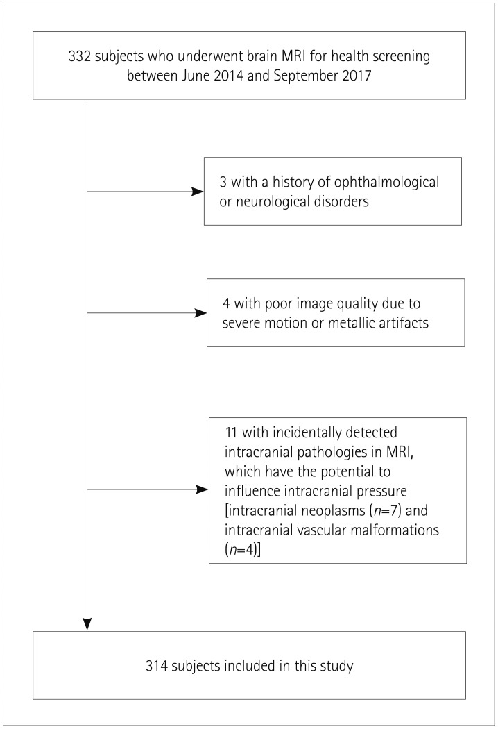
Fig. 1 Flow diagram of subject selection.

Fig. 2 Sample image showing how the ONSD and ETD are estimated. ETD: eyeball transverse diameter, MRA: magnetic resonance angiography, ONSD: optic nerve sheath diameter, TOF: time-of-flight.
Cited by 1 articles
-
Association between optic nerve sheath diameter/eyeball transverse diameter ratio and neurological outcomes in patients with aneurysmal subarachnoid hemorrhage
Jinsung Kim, Hyungoo Shin, Heekyung Lee
J Korean Neurosurg Soc. 2023;66(6):664-671. doi: 10.3340/jkns.2023.0073.
Reference
-
1. Dunn LT. Raised intracranial pressure. J Neurol Neurosurg Psychiatry. 2002; 73(Suppl 1):i23–i27. PMID: 12185258.
Article2. Balestreri M, Czosnyka M, Hutchinson P, Steiner LA, Hiler M, Smielewski P, et al. Impact of intracranial pressure and cerebral perfusion pressure on severe disability and mortality after head injury. Neurocrit Care. 2006; 4:8–13. PMID: 16498188.
Article3. Stevens RD, Shoykhet M, Cadena R. Emergency neurological life support: intracranial hypertension and herniation. Neurocrit Care. 2015; 23(Suppl 2):S76–S82. PMID: 26438459.
Article4. Raboel PH, Bartek J Jr, Andresen M, Bellander BM, Romner B. Intracranial pressure monitoring: invasive versus non-invasive methods-a review. Crit Care Res Pract. 2012; 2012:950393. PMID: 22720148.
Article5. Czosnyka M, Pickard JD. Monitoring and interpretation of intracranial pressure. J Neurol Neurosurg Psychiatry. 2004; 75:813–821. PMID: 15145991.
Article6. Kristiansson H, Nissborg E, Bartek J Jr, Andresen M, Reinstrup P, Romner B. Measuring elevated intracranial pressure through noninvasive methods: a review of the literature. J Neurosurg Anesthesiol. 2013; 25:372–385. PMID: 23715045.7. Dubourg J, Javouhey E, Geeraerts T, Messerer M, Kassai B. Ultrasonography of optic nerve sheath diameter for detection of raised intracranial pressure: a systematic review and meta-analysis. Intensive Care Med. 2011; 37:1059–1068. PMID: 21505900.
Article8. Hansen HC, Helmke K. Validation of the optic nerve sheath response to changing cerebrospinal fluid pressure: ultrasound findings during intrathecal infusion tests. J Neurosurg. 1997; 87:34–40. PMID: 9202262.
Article9. Wang LJ, Yao Y, Feng LS, Wang YZ, Zheng NN, Feng JC, et al. Noninvasive and quantitative intracranial pressure estimation using ultrasonographic measurement of optic nerve sheath diameter. Sci Rep. 2017; 7:42063. PMID: 28169341.
Article10. Geeraerts T, Newcombe VF, Coles JP, Abate MG, Perkes IE, Hutchinson PJ, et al. Use of T2-weighted magnetic resonance imaging of the optic nerve sheath to detect raised intracranial pressure. Crit Care. 2008; 12:R114. PMID: 18786243.
Article11. Mashima Y, Oshitari K, Imamura Y, Momoshima S, Shiga H, Oguchi Y. High-resolution magnetic resonance imaging of the intraorbital optic nerve and subarachnoid space in patients with papilledema and optic atrophy. Arch Ophthalmol. 1996; 114:1197–1203. PMID: 8859078.
Article12. Weigel M, Lagrèze WA, Lazzaro A, Hennig J, Bley TA. Fast and quantitative high-resolution magnetic resonance imaging of the optic nerve at 3.0 tesla. Invest Radiol. 2006; 41:83–86. PMID: 16428977.
Article13. Lagrèze WA, Lazzaro A, Weigel M, Hansen HC, Hennig J, Bley TA. Morphometry of the retrobulbar human optic nerve: comparison between conventional sonography and ultrafast magnetic resonance sequences. Invest Ophthalmol Vis Sci. 2007; 48:1913–1917. PMID: 17460241.14. Bäuerle J, Schuchardt F, Schroeder L, Egger K, Weigel M, Harloff A. Reproducibility and accuracy of optic nerve sheath diameter assessment using ultrasound compared to magnetic resonance imaging. BMC Neurol. 2013; 13:187. PMID: 24289136.
Article15. Kim DH, Jun JS, Kim R. Ultrasonographic measurement of the optic nerve sheath diameter and its association with eyeball transverse diameter in 585 healthy volunteers. Sci Rep. 2017; 7:15906. PMID: 29162911.
Article16. Shirodkar CG, Munta K, Rao SM, Mahesh MU. Correlation of measurement of optic nerve sheath diameter using ultrasound with magnetic resonance imaging. Indian J Crit Care Med. 2015; 19:466–470. PMID: 26321806.17. Tawfik EA, Walker FO, Cartwright MS. Neuromuscular ultrasound of cranial nerves. J Clin Neurol. 2015; 11:109–121. PMID: 25851889.
Article18. Shofty B, Ben-Sira L, Constantini S, Freedman S, Kesler A. Optic nerve sheath diameter on MR imaging: establishment of norms and comparison of pediatric patients with idiopathic intracranial hypertension with healthy controls. AJNR Am J Neuroradiol. 2012; 33:366–369. PMID: 22116116.
Article19. Ko SB. Optic nerve sheath diameter on brain magnetic resonance imaging: a single center study. J Neurocrit Care. 2015; 8:16–24.
Article20. Chen H, Ding GS, Zhao YC, Yu RG, Zhou JX. Ultrasound measurement of optic nerve diameter and optic nerve sheath diameter in healthy Chinese adults. BMC Neurol. 2015; 15:106. PMID: 26148482.
Article21. Rehman H, Khan MS, Nafees M, Rehman AU, Habib A. Optic nerve sheath diameter on sonography in idiopathic intracranial hypertension versus normal. J Coll Physicians Surg Pak. 2016; 26:758–760. PMID: 27671180.22. Wang L, Feng L, Yao Y, Wang Y, Chen Y, Feng J, et al. Optimal optic nerve sheath diameter threshold for the identification of elevated opening pressure on lumbar puncture in a Chinese population. PLoS One. 2015; 10:e0117939. PMID: 25664663.
Article23. Lee SU, Jeon JP, Lee H, Han JH, Seo M, Byoun HS, et al. Optic nerve sheath diameter threshold by ocular ultrasonography for detection of increased intracranial pressure in Korean adult patients with brain lesions. Medicine (Baltimore). 2016; 95:e5061. PMID: 27741121.
Article24. Vaiman M, Gottlieb P, Bekerman I. Quantitative relations between the eyeball, the optic nerve, and the optic canal important for intracranial pressure monitoring. Head Face Med. 2014; 10:32. PMID: 25130267.
Article25. Vaiman M, Sigal T, Kimiagar I, Bekerman I. Intracranial pressure assessment in traumatic head injury with hemorrhage via optic nerve sheath diameter. J Neurotrauma. 2016; 33:2147–2153. PMID: 27048793.
Article26. Vaiman M, Sigal T, Kimiagar I, Bekerman I. Noninvasive assessment of the intracranial pressure in non-traumatic intracranial hemorrhage. J Clin Neurosci. 2016; 34:177–181. PMID: 27612672.
Article27. Bekerman I, Sigal T, Kimiagar I, Ben Ely A, Vaiman M. The quantitative evaluation of intracranial pressure by optic nerve sheath diameter/eye diameter CT measurement. Am J Emerg Med. 2016; 34:2336–2342. PMID: 27717720.
Article28. Wang L, Feng L, Yao Y, Deng F, Wang Y, Feng J, et al. Ultrasonographic evaluation of optic nerve sheath diameter among healthy Chinese adults. Ultrasound Med Biol. 2016; 42:683–688. PMID: 26738627.
Article29. Goeres P, Zeiler FA, Unger B, Karakitsos D, Gillman LM. Ultrasound assessment of optic nerve sheath diameter in healthy volunteers. J Crit Care. 2016; 31:168–171. PMID: 26596508.
Article
- Full Text Links
-
- Actions
-
Cited
- CITED
-
- Close
- Share
-
- Similar articles
-
- Comparison of the effects of normal and low blood pressure regulation on the optic nerve sheath diameter in robot assisted laparoscopic radical prostatectomy
- Optic nerve sheath meningioma mimicking optic perineuritis
- Optic Nerve Sheath Meningocele with Optic Disc Pit
- Optic Neuropathy with Diffusion Weighted High Signal Changes in Optic Nerve
- Ultrasonographic measurement of optic nerve sheath diameter in normal dogs
