J Periodontal Implant Sci.
2018 Jun;48(3):174-181. 10.5051/jpis.2018.48.3.174.
A radiographic evaluation of graft height changes after maxillary sinus augmentation
- Affiliations
-
- 1Department of Periodontology, Wonkwang University Daejeon Dental Hospital, Wonkwang University School of Dentistry, Daejeon, Korea. seongnyum@wonkwang.ac.kr
- KMID: 2414930
- DOI: http://doi.org/10.5051/jpis.2018.48.3.174
Abstract
- PURPOSE
The aims of the present study were to quantitatively assess graft height changes after sinus lift procedures and to analyze the factors that influenced graft height changes, including the residual bone height before surgery, surgical approach, and tooth type.
METHODS
A total of 39 maxillary posterior implants placed during a simultaneous sinus lift procedure were evaluated. Panoramic radiographs of all patients were taken immediately after implant installation and at 3 months, 6 months, 1 year, 2 years, and 3 years. To analyze graft height changes over time, we measured the distance between the implant platform and the base of the grafted sinus floor at 3 locations. The radiographs were analyzed by a single examiner.
RESULTS
Graft height tended to decrease over time, and a statistically significant difference was observed at 2 years compared to baseline (P < 0.05). There was no statistically significant difference in graft height change according to the surgical approach or tooth type. For residual bone height, a statistically significant difference in graft height change was found between those with 4-7 mm of residual bone height and those with ≥7 mm (P < 0.05).
CONCLUSIONS
Graft height after sinus lift procedures significantly decreased at 2 years compared to baseline after sinus augmentation. Further studies should be done with controlled variables, and prospective studies with 3-dimensional images are needed to clarify the factors that influence graft height changes.
MeSH Terms
Figure
-
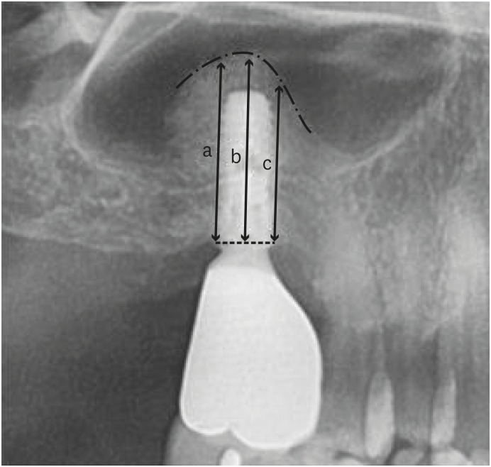
Figure 1 Radiographic measurements of graft height change using a panoramic radiograph. Lower dotted line and upper dotted line mean implant platform and grafted sinus floor, respectively.a: The distance from the implant platform to the base of grafted sinus floor at the mesial side of the implant, b: The distance from the implant platform to the base of the grafted sinus floor at the middle side of the implant, c: The distance from the implant platform to the base of the grafted sinus floor at the distal side of the implant.
Reference
-
1. Davarpanah M, Martinez H, Tecucianu JF, Hage G, Lazzara R. The modified osteotome technique. Int J Periodontics Restorative Dent. 2001; 21:599–607. PMID: 11794571.2. Tatum H Jr. Maxillary and sinus implant reconstructions. Dent Clin North Am. 1986; 30:207–229. PMID: 3516738.3. Boyne PJ, James RA. Grafting of the maxillary sinus floor with autogenous marrow and bone. J Oral Surg. 1980; 38:613–616. PMID: 6993637.4. Summers RB. A new concept in maxillary implant surgery: the osteotome technique. Compendium. 1994; 15:152–156. PMID: 8055503.5. Summers RB. The osteotome technique: Part 3--Less invasive methods of elevating the sinus floor. Compendium. 1994; 15:698–710. PMID: 7994726.6. Wang HL, Katranji A. ABC sinus augmentation classification. Int J Periodontics Restorative Dent. 2008; 28:383–389. PMID: 18717377.7. Wanschitz F, Figl M, Wagner A, Rolf E. Measurement of volume changes after sinus floor augmentation with a phycogenic hydroxyapatite. Int J Oral Maxillofac Implants. 2006; 21:433–438. PMID: 16796287.8. Hatano N, Shimizu Y, Ooya K. A clinical long-term radiographic evaluation of graft height changes after maxillary sinus floor augmentation with a 2:1 autogenous bone/xenograft mixture and simultaneous placement of dental implants. Clin Oral Implants Res. 2004; 15:339–345. PMID: 15142097.9. van den Bergh JP, ten Bruggenkate CM, Disch FJ, Tuinzing DB. Anatomical aspects of sinus floor elevations. Clin Oral Implants Res. 2000; 11:256–265. PMID: 11168217.
Article10. Velloso GR, Vidigal GM Jr, de Freitas MM, Garcia de Brito OF, Manso MC, Groisman M. Tridimensional analysis of maxillary sinus anatomy related to sinus lift procedure. Implant Dent. 2006; 15:192–196. PMID: 16766903.
Article11. Chan HL, Suarez F, Monje A, Benavides E, Wang HL. Evaluation of maxillary sinus width on cone-beam computed tomography for sinus augmentation and new sinus classification based on sinus width. Clin Oral Implants Res. 2014; 25:647–652. PMID: 23043676.
Article12. Wen SC, Chan HL, Wang HL. Classification and management of antral septa for maxillary sinus augmentation. Int J Periodontics Restorative Dent. 2013; 33:509–517. PMID: 23820711.
Article13. Kent JN, Block MS. Simultaneous maxillary sinus floor bone grafting and placement of hydroxylapatite-coated implants. J Oral Maxillofac Surg. 1989; 47:238–242. PMID: 2646403.
Article14. Hieu PD, Chung JH, Yim SB, Hong KS. A radiographical study on the changes in height of grafting materials after sinus lift: a comparison between two types of xenogenic materials. J Periodontal Implant Sci. 2010; 40:25–32. PMID: 20498756.
Article15. Alayan J, Ivanovski S. A prospective controlled trial comparing xenograft/autogenous bone and collagen-stabilized xenograft for maxillary sinus augmentation-complications, patient-reported outcomes and volumetric analysis. Clin Oral Implants Res. 2018; 29:248–262. PMID: 29231263.
Article16. Pereira RS, Gorla LF, Boos FB, Okamoto R, Garcia Júnior IR, Hochuli-Vieira E. Use of autogenous bone and beta-tricalcium phosphate in maxillary sinus lifting: histomorphometric study and immunohistochemical assessment of RUNX2 and VEGF. Int J Oral Maxillofac Surg. 2017; 46:503–510. PMID: 28185708.
Article17. Chanavaz M. Maxillary sinus: anatomy, physiology, surgery, and bone grafting related to implantology--eleven years of surgical experience (1979–1990). J Oral Implantol. 1990; 16:199–209. PMID: 2098563.18. Hürzeler MB, Kirsch A, Ackermann KL, Quiñones CR. Reconstruction of the severely resorbed maxilla with dental implants in the augmented maxillary sinus: a 5-year clinical investigation. Int J Oral Maxillofac Implants. 1996; 11:466–475. PMID: 8803342.19. Listrom RD, Symington JM. Osseointegrated dental implants in conjunction with bone grafts. Int J Oral Maxillofac Surg. 1988; 17:116–118. PMID: 3133419.
Article20. Geurs NC, Wang IC, Shulman LB, Jeffcoat MK. Retrospective radiographic analysis of sinus graft and implant placement procedures from the Academy of Osseointegration Consensus Conference on Sinus Grafts. Int J Periodontics Restorative Dent. 2001; 21:517–523. PMID: 11693244.21. Cho SH, Kim OS. Radiographic change of grafted sinus floor after maxillary sinus floor elevation and placement of dental implant. J Korean Acad Periodontol. 2006; 36:345–359.
Article22. McCaul LK, Jenkins WM, Kay EJ. The reasons for the extraction of various tooth types in Scotland: a 15-year follow up. J Dent. 2001; 29:401–407. PMID: 11520588.
Article23. Fredholm U, Bolin A, Andersson L. Preimplant radiographic assessment of available maxillary bone support. Comparison of tomographic and panoramic technique. Swed Dent J. 1993; 17:103–109. PMID: 8356535.
- Full Text Links
-
- Actions
-
Cited
- CITED
-
- Close
- Share
-
- Similar articles
-
- Case series of maxillary sinus augmentation with biphasic calcium phosphate: a clinical and radiographic study
- A Radiographic Evaluation of Graft Height Changes after Maxillary Sinus Augmentation according to Single-unit Implants and Multi-unit Implants
- A radiographic evaluation of graft height changes after maxillary sinus augmentation and placement of dental implants
- One-staged sinus bone graft using tapered porous surfaced implant in lower residual bone height of maxillary sinus
- A Case of Maxillary Sinusitis after Sinus Floor Augmentation
