Korean Circ J.
2018 Jul;48(7):658-660. 10.4070/kcj.2018.0019.
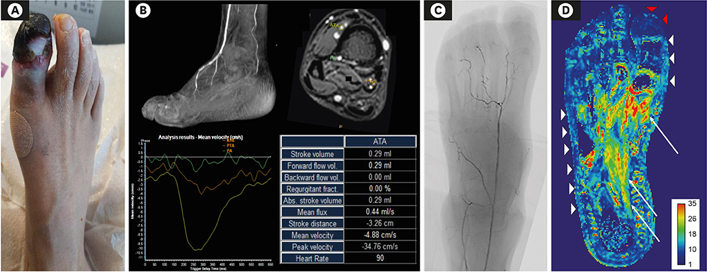
Assessment of Tissue Perfusion with Blood Oxygenation Level-Dependent Magnetic Resonance Imaging in Critical Limb Ischemia
- Affiliations
-
- 1Department of Radiology and Research Institute of Radiology, Asan Medical Center, University of Ulsan College of Medicine, Seoul, Korea.
- 2Division of Cardiology, Department of Internal Medicine, Asan Medical Center, University of Ulsan College of Medicine, Seoul, Korea. pilmo11@hanmail.net
- 3Division of Endocrinology, Department of Internal Medicine, Asan Medical Center, University of Ulsan College of Medicine, Seoul, Korea.
- KMID: 2414902
- DOI: http://doi.org/10.4070/kcj.2018.0019
Abstract
- No abstract available.
Figure
Reference
-
1. Stacy MR, Qiu M, Papademetris X, Caracciolo CM, Constable RT, Sinusas AJ. Application of BOLD magnetic resonance imaging for evaluating regional volumetric foot tissue oxygenation: a feasibility study in healthy volunteers. Eur J Vasc Endovasc Surg. 2016; 51:743–749.
Article2. Bajwa A, Wesolowski R, Patel A, et al. Blood oxygenation level-dependent CMR-derived measures in critical limb ischemia and changes with revascularization. J Am Coll Cardiol. 2016; 67:420–431.3. Ledermann HP, Heidecker HG, Schulte AC, et al. Calf muscles imaged at BOLD MR: correlation with TcPO2 and flowmetry measurements during ischemia and reactive hyperemia--initial experience. Radiology. 2006; 241:477–484.4. Bajwa A, Wesolowski R, Patel A, et al. Assessment of tissue perfusion in the lower limb: current methods and techniques under development. Circ Cardiovasc Imaging. 2014; 7:836–843.
- Full Text Links
- Actions
-
Cited
- CITED
-
- Close
- Share
- Similar articles
-
- Evaluation Methods for Foot Perfusion in Critical Limb Ischemia
- Assessment of Myocardial Ischemia Using Stress Perfusion Cardiovascular Magnetic Resonance
- Functional Magnetic Resonance Imaging with Arterial Spin Labeling: Techniques and Potential Clinical and Research Applications
- Safety and Efficacy of Distal Perfusion Catheterization to Prevent Limb Ischemia after Common Femoral Artery Cannulation for Extracorporeal Membrane Oxygenation
- Spinal Cord Infarction in a Patient Undergoing Veno-arterial Extracorporeal Membrane Oxygenation


