Ann Dermatol.
2018 Apr;30(2):241-242. 10.5021/ad.2018.30.2.241.
Tinea Faciei in a Mother and Daughter Caused by Arthroderma benhamiae
- Affiliations
-
- 1Department of Dermatology, Kyungpook National University School of Medicine, Daegu, Korea. weonju@knu.ac.kr
- 2Institute of Medical Mycology, Catholic Skin Clinic, Daegu, Korea.
- KMID: 2414693
- DOI: http://doi.org/10.5021/ad.2018.30.2.241
Abstract
- No abstract available.
Figure
-
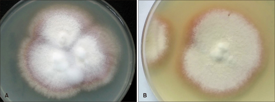
Fig. 1 (A) Peripherally radiating and centrally raised, granular and downy colonies cultured from mother and (B) her daughter.
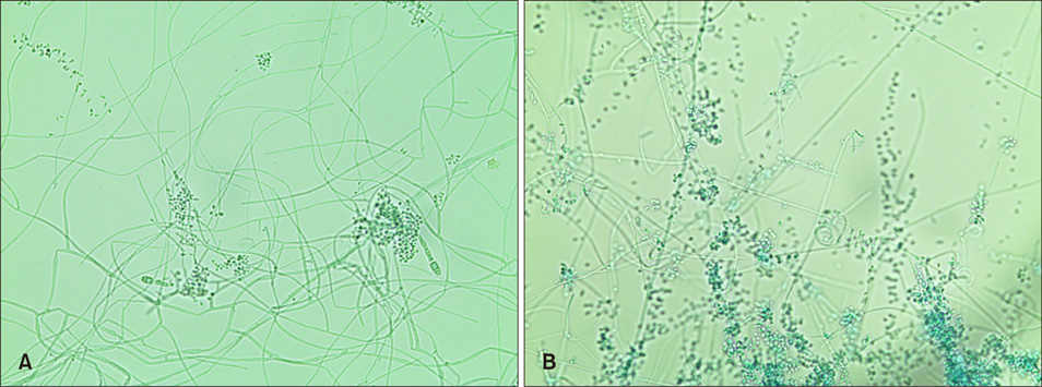
Fig. 2 (A) Septate mycelium with a great number of small round microconidia and a few macroconidia from mother. (B) Septate mycelium with a great number of small round microconidia and spiral hyphae from her daughter (A, B: lactophenol cotton blue stain, ×200).
Reference
-
1. Kano R, Nakamura Y, Yasuda K, Watari T, Watanabe S, Takahashi H, et al. The first isolation of Arthroderma benhamiae in Japan. Microbiol Immunol. 1998; 42:575–578.2. Budihardja D, Freund V, Mayser P. Widespread erosive tinea corporis by Arthroderma benhamiae in a renal transplant recipient: case report. Mycoses. 2010; 53:530–532.
Article3. Jun JB, Sang YH, Chung SL, Choi JS, Suh SB. The mycological and molecular biological studies on Arthroderma benhamiae isolated for the first time in Korea. Korean J Med Mycol. 2004; 9:12–27.4. Mayser P, Budihardja D. A simple and rapid method to differentiate Arthroderma benhamiae from Microsporum canis. J Dtsch Dermatol Ges. 2013; 11:322–327.
Article5. Nenoff P, Uhrlaß S, Krüger C, Erhard M, Hipler UC, Seyfarth F, et al. Trichophyton species of Arthroderma benhamiae-a new infectious agent in dermatology. J Dtsch Dermatol Ges. 2014; 12:571–581.
Article
- Full Text Links
-
- Actions
-
Cited
- CITED
-
- Close
- Share
-
- Similar articles
-
- Mycologic Findings of Trichophyton mentagrophytes var. mentagrophytes Isolated from the Patients with Dermatophytosis in Taegu Area and Microsporum Persicolor
- A Case of Tinea Faciei Caused by Trichophyton mentagrophytes with Atypical Presentation
- The Clinical and Mycological Study of Tinea Faciei in Daegu
- The Mycological and Molecular Biological Studies on Arthroderma benhamiae Isolated for the First Time in Korea
- Two cases of tinea faciei with atypical clinical manifestation
