Breast Cancer Detection in a Screening Population: Comparison of Digital Mammography, Computer-Aided Detection Applied to Digital Mammography and Breast Ultrasound
- Affiliations
-
- 1Department of Radiology, Korea University Anam Hospital, Korea University College of Medicine, Seoul, Korea.
- 2Department of Radiology, Korea University Ansan Hospital, Korea University College of Medicine, Ansan, Korea. seoboky@korea.ac.kr
- 3Department of Radiology, Korea University Guro Hospital, Korea University College of Medicine, Seoul, Korea.
- 4Department of Mathematics, School of Natural Sciences, Hanyang University, Seoul, Korea.
- KMID: 2413957
- DOI: http://doi.org/10.4048/jbc.2016.19.3.316
Abstract
- PURPOSE
We aimed to compare the detection of breast cancer using full-field digital mammography (FFDM), FFDM with computer-aided detection (FFDM+CAD), ultrasound (US), and FFDM+CAD plus US (FFDM+CAD+US), and to investigate the factors affecting cancer detection.
METHODS
In this retrospective study conducted from 2008 to 2012, 48,251 women underwent FFDM and US for cancer screening. One hundred seventy-one breast cancers were detected: 115 invasive cancers and 56 carcinomas in situ. Two radiologists evaluated the imaging findings of FFDM, FFDM+CAD, and US, based on the Breast Imaging Reporting and Data System lexicon of the American College of Radiology by consensus. We reviewed the clinical and the pathological data to investigate factors affecting cancer detection. We statistically used generalized estimation equations with a logit link to compare the cancer detectability of different imaging modalities. To compare the various factors affecting detection versus nondetection, we used Wilcoxon rank sum, chi-square, or Fisher exact test.
RESULTS
The detectability of breast cancer by US (96.5%) or FFDM+CAD+US (100%) was superior to that of FFDM (87.1%) (p=0.019 or p<0.001, respectively) or FFDM+ CAD (88.3%) (p=0.050 or p<0.001, respectively). However, cancer detectability was not significantly different between FFDM versus FFDM+CAD (p=1.000) and US alone versus FFDM+CAD+US (p=0.126). The tumor size influenced cancer detectability by all imaging modalities (p<0.050). In FFDM and FFDM+CAD, the nondetecting group consisted of younger patients and patients with a denser breast composition (p<0.050). In breast US, carcinoma in situ was more frequent in the nondetecting group (p=0.014).
CONCLUSION
For breast cancer screening, breast US alone is satisfactory for all age groups, although FFDM+ CAD+US is the perfect screening method. Patient age, breast composition, and pathological tumor size and type may influence cancer detection during screening.
Keyword
MeSH Terms
Figure
-
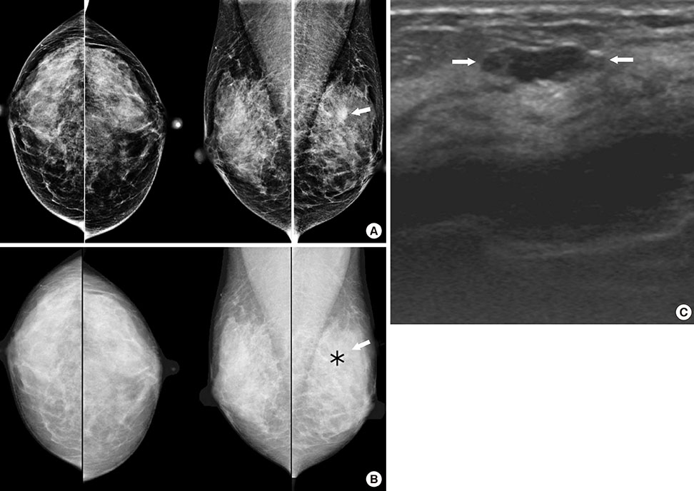
Figure 1 Mammographic and ultrasound findings of a 35-year-old woman with ductal carcinomas in situ (DCIS). Screening mammography (A) demonstrated a focal asymmetry in the left upper breast (arrow) and computer-aided detection applied mammography (B) detected the lesion (asterisk, marked by computer-aided detection program) (arrow). The breast ultrasound (C) demonstrated a ductal change in the left upper breast (arrows); this was a pathologically proven DCIS.
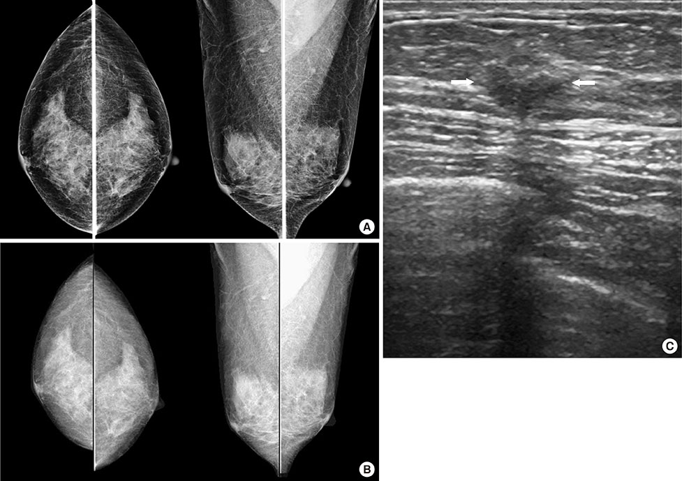
Figure 2 Mammographic and ultrasound findings of a 46-year-old woman with invasive ductal carcinoma. Screening mammography (A) and computer-aided detection applied mammography (B) did not show any abnormal findings. The breast ultrasound (C) demonstrated an indistinct oval hypoechoic mass in the left upper outer quadrant (arrows); this was a pathologically verified cancer.
Reference
-
1. Pisano ED, Yaffe MJ. Digital mammography. Radiology. 2005; 234:353–362.
Article2. Shtern F. Digital mammography and related technologies: a perspective from the National Cancer Institute. Radiology. 1992; 183:629–630.
Article3. Pisano ED, Gatsonis CA, Yaffe MJ, Hendrick RE, Tosteson AN, Fryback DG, et al. American College of Radiology Imaging Network digital mammographic imaging screening trial: objectives and methodology. Radiology. 2005; 236:404–412.
Article4. Pisano ED, Gatsonis C, Hendrick E, Yaffe M, Baum JK, Acharyya S, et al. Diagnostic performance of digital versus film mammography for breast-cancer screening. N Engl J Med. 2005; 353:1773–1783.
Article5. Brem RF, Baum J, Lechner M, Kaplan S, Souders S, Naul LG, et al. Improvement in sensitivity of screening mammography with computer-aided detection: a multiinstitutional trial. AJR Am J Roentgenol. 2003; 181:687–693.
Article6. Warren Burhenne LJ, Wood SA, D'Orsi CJ, Feig SA, Kopans DB, O'Shaughnessy KF, et al. Potential contribution of computer-aided detection to the sensitivity of screening mammography. Radiology. 2000; 215:554–562.
Article7. Dean JC, Ilvento CC. Improved cancer detection using computer-aided detection with diagnostic and screening mammography: prospective study of 104 cancers. AJR Am J Roentgenol. 2006; 187:20–28.
Article8. Berg WA, Gilbreath PL. Multicentric and multifocal cancer: whole-breast US in preoperative evaluation. Radiology. 2000; 214:59–66.
Article9. Crystal P, Strano SD, Shcharynski S, Koretz MJ. Using sonography to screen women with mammographically dense breasts. AJR Am J Roentgenol. 2003; 181:177–182.
Article10. Kaplan SS. Clinical utility of bilateral whole-breast US in the evaluation of women with dense breast tissue. Radiology. 2001; 221:641–649.
Article11. Leconte I, Feger C, Galant C, Berlière M, Berg BV, D'Hoore W, et al. Mammography and subsequent whole-breast sonography of nonpalpable breast cancers: the importance of radiologic breast density. AJR Am J Roentgenol. 2003; 180:1675–1679.
Article12. D'Orsi CJ, Sickles EA, Mendelson EB, Morris EA. ACR BI-RADS Atlas: Breast Imaging Reporting and Data System. Reston: American College of Radiology;2013.13. Liang KY, Zeger SL. Longitudinal data analysis using generalized linear models. Biometrika. 1986; 73:13–22.
Article14. Nyström L, Rutqvist LE, Wall S, Lindgren A, Lindqvist M, Rydén S, et al. Breast cancer screening with mammography: overview of Swedish randomised trials. Lancet. 1993; 341:973–978.
Article15. Tabar L, Fagerberg G, Chen HH, Duffy SW, Smart CR, Gad A, et al. Efficacy of breast cancer screening by age: new results from the Swedish two-county trial. Cancer. 1995; 75:2507–2517.
Article16. Mandelson MT, Oestreicher N, Porter PL, White D, Finder CA, Taplin SH, et al. Breast density as a predictor of mammographic detection: comparison of interval- and screen-detected cancers. J Natl Cancer Inst. 2000; 92:1081–1087.
Article17. Kim SJ, Moon WK, Cho N, Cha JH, Kim SM, Im JG. Computer-aided detection in full-field digital mammography: sensitivity and reproducibility in serial examinations. Radiology. 2008; 246:71–80.
Article18. van den Biggelaar FJ, Kessels AG, van Engelshoven JM, Boetes C, Flobbe K. Computer-aided detection in full-field digital mammography in a clinical population: performance of radiologist and technologists. Breast Cancer Res Treat. 2010; 120:499–506.
Article19. Bolivar AV, Gomez SS, Merino P, Alonso-Bartolomé P, Garcia EO, Cacho PM, et al. Computer-aided detection system applied to full-field digital mammograms. Acta Radiol. 2010; 51:1086–1092.
Article20. Sadaf A, Crystal P, Scaranelo A, Helbich T. Performance of computer-aided detection applied to full-field digital mammography in detection of breast cancers. Eur J Radiol. 2011; 77:457–461.
Article21. Murakami R, Kumita S, Tani H, Yoshida T, Sugizaki K, Kuwako T, et al. Detection of breast cancer with a computer-aided detection applied to full-field digital mammography. J Digit Imaging. 2013; 26:768–773.
Article22. Korpraphong P, Limsuwarn P, Tangcharoensathien W, Ansusingha T, Thephamongkhol K, Chuthapisith S. Improving breast cancer detection using ultrasonography in asymptomatic women with non-fatty breast density. Acta Radiol. 2014; 55:903–908.
Article23. Yang SK, Moon WK, Cho N, Park JS, Cha JH, Kim SM, et al. Screening mammography-detected cancers: sensitivity of a computer-aided detection system applied to full-field digital mammograms. Radiology. 2007; 244:104–111.
Article24. Kim SH, Kim MH, Oh KK. Analysis and comparison of breast density according to age on mammogram between Korean and Western women. J Korean Radiol Soc. 2000; 42:1009–1014.
Article25. Kim HY, Seo BK, Kim HY, Yie A, Cho KR, Seol HY, et al. Additional breast ultrasound examinations in clustered calcifications: for improving diagnostic performance. J Breast Cancer. 2009; 12:142–150.
Article26. Barreau B, de Mascarel I, Feuga C, MacGrogan G, Dilhuydy MH, Picot V, et al. Mammography of ductal carcinoma in situ of the breast: review of 909 cases with radiographic-pathologic correlations. Eur J Radiol. 2005; 54:55–61.
Article27. Holland R, Peterse JL, Millis RR, Eusebi V, Faverly D, van de Vijver MJ, et al. Ductal carcinoma in situ: a proposal for a new classification. Semin Diagn Pathol. 1994; 11:167–180.28. Jin ZQ, Lin MY, Hao WQ, Jiang HT, Zhang L, Hu WH, et al. Diagnostic evaluation of ductal carcinoma in situ of the breast: ultrasonographic, mammographic and histopathologic correlations. Ultrasound Med Biol. 2015; 41:47–55.
Article29. Izumori A, Takebe K, Sato A. Ultrasound findings and histological features of ductal carcinoma in situ detected by ultrasound examination alone. Breast Cancer. 2010; 17:136–141.
Article
- Full Text Links
-
- Actions
-
Cited
- CITED
-
- Close
- Share
-
- Similar articles
-
- Digital Mammography
- New Trends in Breast Imaging
- Applications of Artificial Intelligence in Mammography from a Development and Validation Perspective
- Digital Mammography as a Screening Tool in Korea
- 3D Computer-Aided Detection for Digital Breast Tomosynthesis: Comparison with 2D Computer-Aided Detection for Digital Mammography in the Detection of Calcifications
