Reliability of Breast Ultrasound BI-RADS Final Assessment in Mammographically Negative Patients with Nipple Discharge and Radiologic Predictors of Malignancy
- Affiliations
-
- 1Department of Radiology, Breast Cancer Clinic, Severance Hospital, Research Institute of Radiological Science, Yonsei University College of Medicine, Seoul, Korea. mines@yuhs.ac
- KMID: 2413956
- DOI: http://doi.org/10.4048/jbc.2016.19.3.308
Abstract
- PURPOSE
The purpose of this study was to retrospectively investigate the reliability of breast ultrasound (US) Breast Imaging Reporting and Data System (BI-RADS) final assessment in mammographically negative patients with pathologic nipple discharge, and to determine the clinical and ultrasonographic variables associated with malignancy in this group of patients.
METHODS
A total of 65 patients with 67 mammographically negative breast lesions that were pathologically confirmed through US-guided biopsy were included.
RESULTS
Of the 53 BI-RADS category 4 and 5 lesions, eight (15.1%) were malignant (six ductal carcinomas in situ, one invasive ductal carcinoma, and one solid papillary carcinoma). There was no malignancy among the remaining 14 category 3 lesions. Malignant lesions more frequently displayed a round or irregular shape (75.0%, 6/8; p=0.030) and nonparallel orientation (33.3%, 4/12; p=0.029) compared to the benign lesions. The increase in the BI-RADS category corresponded with a rise in the malignancy rate (p=0.004).
CONCLUSION
The BI-RADS lexicon and final assessment of breast US reliably detect and characterize malignancy in mammographically negative patients with pathologic nipple discharge.
Keyword
MeSH Terms
Figure
-

Figure 1 Flow chart of the study investigating the reliability of breast ultrasound Breast Imaging Reporting and Data System (BI-RADS) final assessment in mammographically negative patients with nipple discharge and radiologic predictors of malignancy. US=ultrasound.
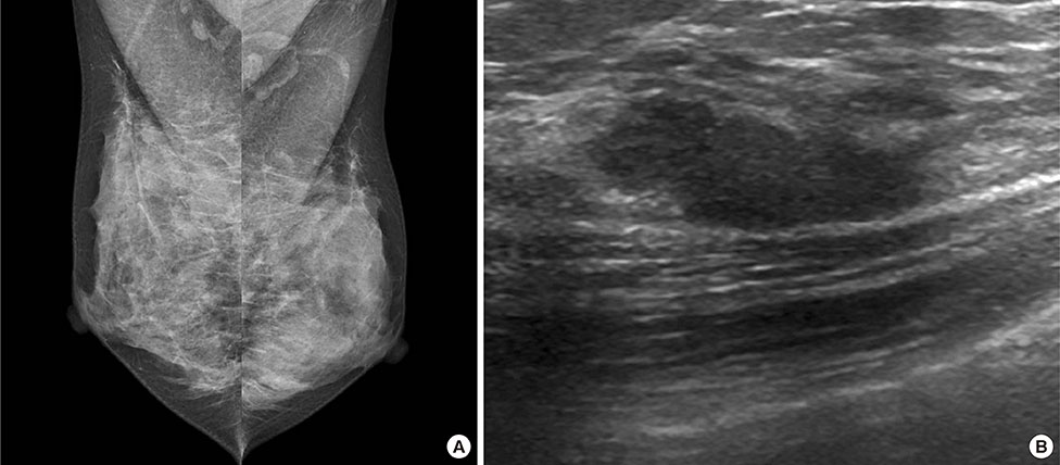
Figure 2 Radiologic findings of a 38-year-old woman with a several-week history of spontaneous serous discharge from the right nipple. (A) Mediolateral mammography of the both breast shows dense breasts with no abnormalities to explain right nipple discharge (Breast Imaging Reporting and Data System [BI-RADS] category 1). (B) Longitudinal ultrasound images show a 2.0 cm sized isoechoic, oval shaped, microlobulated mass with parallel orientation which was located at 1 o'clock 2 cm from nipple (BI-RADS category 3). Ultrasound-guided vacuum-assisted biopsy was performed and the pathologic result was consistent with fibrocystic disease with stromal fibrosis. After the procedure, the patient had no persistent nipple discharge, and no malignancy was detected during a 2-year follow-up period.
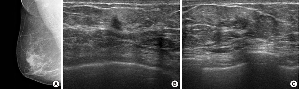
Figure 3 Radiologic findings of a 73-year-old woman with a 1-week history of spontaneous bloody discharge from the right nipple. (A) Mediolateral mammography of right breast shows no definite abnormality to explain right bloody nipple discharge. Transverse (B) and longitudinal (C) ultrasound images of the right breast show a 1.0 cm sized isoechoic, irregularly shaped, spiculated mass with nonparallel orientation (Breast Imaging Reporting and Data System category 5) which was located at 8 o'clock 1 cm from nipple. Ultrasound-guided biopsy was performed, and the pathologic result was consistent with ductal carcinoma in situ. Subsequent surgery was performed.
Reference
-
1. Leis HP Jr. Management of nipple discharge. World J Surg. 1989; 13:736–742.
Article2. Leis HP Jr, Greene FL, Cammarata A, Hilfer SE. Nipple discharge: surgical significance. South Med J. 1988; 81:20–26.
Article3. Gülay H, Bora S, Kìlìçturgay S, Hamaloğlu E, Göksel HA. Management of nipple discharge. J Am Coll Surg. 1994; 178:471–474.4. Morrogh M, Park A, Elkin EB, King TA. Lessons learned from 416 cases of nipple discharge of the breast. Am J Surg. 2010; 200:73–80.
Article5. Rissanen T, Reinikainen H, Apaja-Sarkkinen M. Breast sonography in localizing the cause of nipple discharge: comparison with galactography in 52 patients. J Ultrasound Med. 2007; 26:1031–1039.6. Alcock C, Layer GT. Predicting occult malignancy in nipple discharge. ANZ J Surg. 2010; 80:646–649.
Article7. King TA, Carter KM, Bolton JS, Fuhrman GM. A simple approach to nipple discharge. Am Surg. 2000; 66:960–965.8. Simmons R, Adamovich T, Brennan M, Christos P, Schultz M, Eisen C, et al. Nonsurgical evaluation of pathologic nipple discharge. Ann Surg Oncol. 2003; 10:113–116.
Article9. Dawes LG, Bowen C, Venta LA, Morrow M. Ductography for nipple discharge: no replacement for ductal excision. Surgery. 1998; 124:685–691.
Article10. Gioffrè Florio M, Manganaro T, Pollicino A, Scarfo P, Micali B. Surgical approach to nipple discharge: a ten-year experience. J Surg Oncol. 1999; 71:235–238.
Article11. Murad TM, Contesso G, Mouriesse H. Nipple discharge from the breast. Ann Surg. 1982; 195:259–264.
Article12. Vargas HI, Romero L, Chlebowski RT. Management of bloody nipple discharge. Curr Treat Options Oncol. 2002; 3:157–161.
Article13. Leconte I, Feger C, Galant C, Berlière M, Berg BV, D'Hoore W, et al. Mammography and subsequent whole-breast sonography of nonpalpable breast cancers: the importance of radiologic breast density. AJR Am J Roentgenol. 2003; 180:1675–1679.
Article14. Kim EK, Ko KH, Oh KK, Kwak JY, You JK, Kim MJ, et al. Clinical application of the BI-RADS final assessment to breast sonography in conjunction with mammography. AJR Am J Roentgenol. 2008; 190:1209–1215.
Article15. D'Orsi CJ, Sickles EA, Mendelson EB, Morris EA. ACR BI-RADS Atlas: Breast Imaging Reporting and Data System. Reston: American College of Radiology;2013.16. Kim MJ, Kim EK, Kwak JY, Son EJ, Park BW, Kim SI, et al. Nonmalignant papillary lesions of the breast at US-guided directional vacuumassisted removal: a preliminary report. Eur Radiol. 2008; 18:1774–1783.
Article17. Kim WH, Chang JM, Moon WK, Cho N, Yi A, Koo HR, et al. Intraductal mass on breast ultrasound: final outcomes and predictors of malignancy. AJR Am J Roentgenol. 2013; 200:932–937.
Article18. Ballesio L, Maggi C, Savelli S, Angeletti M, Rabuffi P, Manganaro L, et al. Adjunctive diagnostic value of ultrasonography evaluation in patients with suspected ductal breast disease. Radiol Med. 2007; 112:354–365.
Article19. Bahl M, Baker JA, Greenup RA, Ghate SV. Diagnostic value of ultrasound in female patients with nipple discharge. AJR Am J Roentgenol. 2015; 205:203–208.
Article20. Berg WA, Blume JD, Cormack JB, Mendelson EB, Lehrer D, Böhm-Vélez M, et al. Combined screening with ultrasound and mammography vs mammography alone in women at elevated risk of breast cancer. JAMA. 2008; 299:2151–2163.
Article21. Berg WA, Zhang Z, Lehrer D, Jong RA, Pisano ED, Barr RG, et al. Detection of breast cancer with addition of annual screening ultrasound or a single screening MRI to mammography in women with elevated breast cancer risk. JAMA. 2012; 307:1394–1404.
Article22. Nakahara H, Namba K, Watanabe R, Furusawa H, Matsu T, Akiyama F, et al. A comparison of MR imaging, galactography and ultrasonography in patients with nipple discharge. Breast Cancer. 2003; 10:320–329.
Article23. Adepoju LJ, Chun J, El-Tamer M, Ditkoff BA, Schnabel F, Joseph KA. The value of clinical characteristics and breast-imaging studies in predicting a histopathologic diagnosis of cancer or high-risk lesion in patients with spontaneous nipple discharge. Am J Surg. 2005; 190:644–646.
Article24. Cabioglu N, Hunt KK, Singletary SE, Stephens TW, Marcy S, Meric F, et al. Surgical decision making and factors determining a diagnosis of breast carcinoma in women presenting with nipple discharge. J Am Coll Surg. 2003; 196:354–364.
Article25. Mercado CL. BI-RADS update. Radiol Clin North Am. 2014; 52:481–487.
Article26. Stavros AT, Thickman D, Rapp CL, Dennis MA, Parker SH, Sisney GA. Solid breast nodules: use of sonography to distinguish between benign and malignant lesions. Radiology. 1995; 196:123–134.
Article27. Yoon H, Yoon JH, Kim EK, Moon HJ, Park BW, Kim MJ. Adding ultrasound to the evaluation of patients with pathologic nipple discharge to diagnose additional breast cancers: preliminary data. Ultrasound Med Biol. 2015; 41:2099–2107.
Article
- Full Text Links
-
- Actions
-
Cited
- CITED
-
- Close
- Share
-
- Similar articles
-
- Usefulness of ultrasound elastography in reducing the number of Breast Imaging Reporting and Data System category 3 lesions on ultrasonography
- Breast Imaging Reporting and Data System (BI-RADS): Advantages and Limitations
- Categorization and Evaluation of Usefulness of Breast Lesions with using Ultrasound BI-RADS (Breast Imaging Reporting and Data system)
- Outcomes of US BI-RADS 4A, 4B, and 4C Lesions
- Complex Cysts of the Breast: Analysis using Sonographic Breast Imaging Reporting and Data System
