Ultrasonographic Findings of Subcutaneous Angioleiomyomas in the Extremities Based on Pathologic Subtypes
- Affiliations
-
- 1Department of Radiology, College of Medicine, Inje University Busan Paik Hospital, Busan 47392, Korea. sunjulee98@naver.com
- 2Department of Radiology, College of Medicine, Soonchunhyang University Bucheon Hospital, Bucheon 14584, Korea.
- 3Department of Radiology, Sungkyunkwan University School of Medicine, Kangbuk Samsung Hospital, Seoul 03181, Korea.
- 4Department of Radiology, Dongcheon Dongkang Hospital, Ulsan 44495, Korea.
- 5Department of Radiology, Daegu Fatima Hospital, Daegu 41199, Korea.
- 6Department of Pathology, College of Medicine, Inje University Busan Paik Hospital, Busan 47392, Korea.
- KMID: 2413704
- DOI: http://doi.org/10.3348/kjr.2018.19.4.752
Abstract
OBJECTIVE
The purpose of this study was to describe the ultrasonographic findings of angioleiomyoma based on pathological subtypes.
MATERIALS AND METHODS
Thirty-nine patients with subcutaneous angioleiomyomas in the extremities were retrospectively reviewed by two radiologists and a pathologist. Sonographic images were analyzed to evaluate each tumor's anatomic location, size, shape, margin, heterogeneity, echogenicity, associated findings, and vascularity.
RESULTS
Angioleiomyomas were divided into 3 subtypes: capillary (n = 16), venous (n = 22), and cavernous (n = 1). The one cavernous angioleiomyoma was a hypoechoic mass with rich vascularity. Hypoechogenicity was more frequently observed for venous tumors (77.3%) than for capillary tumors (43.8%), and isoechogenicity was more frequently observed for capillary tumors (56.2%) than for venous tumors (22.7%). Moderate vascularity was more frequently observed for venous tumors (59.1%) than for capillary tumors (12.5%), and little vascularity was more frequently observed for capillary tumors (62.5%) than for venous tumors (13.6%). The aforementioned findings including echogenicity (p = 0.034) and vascularity (p = 0.003) were statistically significant.
CONCLUSION
Awareness of sonographic findings of angioleiomyomas based on pathologic subtypes could be helpful for diagnosing angioleiomyoma and could increase diagnostic accuracy for superficial soft-tissue masses in our practice.
MeSH Terms
Figure
-
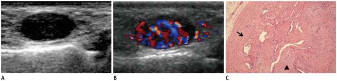
Fig. 1 35-year-old female with palpable mass that was confirmed to be cavernous angioleiomyoma in 1st web space of left hand.A. Gray-scale sonogram in longitudinal plane shows well-defined, homogeneously hypoechoic mass. B. Color Doppler sonogram of same lesion shows abundant vascularity with several feeding vessels. C. Photomicrograph shows that this mass had dilated vascular channels with minimal smooth muscle that merged with surrounding smooth muscles (arrow) and was mixed with thick-walled vessels (arrow head), indicating that tumor was cavernous angioleiomyoma (original magnification, × 100, H&E).
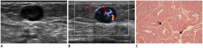
Fig. 2 67-year-old female with palpable mass that was confirmed to be venous angioleiomyoma in 4th–5th palmar region of hand.A. Gray-scale sonogram in longitudinal plane shows well-defined, homogeneously hypoechoic mass. B. Color Doppler sonogram of same lesion shows moderate vascularity with several feeding vessels. C. Photomicrograph shows that this mass had vessels (arrows) with thick muscular walls that merged with smooth muscle bundles, indicating that tumor was venous angioleiomyoma (original magnification, × 100, H&E).
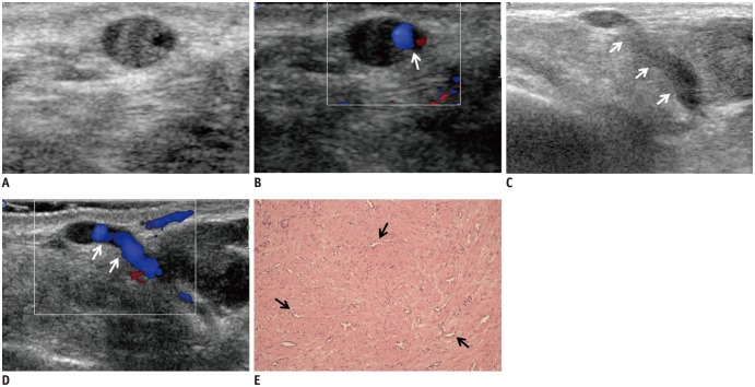
Fig. 3 53-year-old female with palpable mass that was confirmed to be capillary angioleiomyoma in left calf.A, B. Transverse scan of gray-scale and color Doppler sonogram show well-defined, homogeneously isoechoic mass with little vascularity and feeding vessel (arrow). C, D. Longitudinal scan of gray-scale and color Doppler sonogram show absence of vascularity in mass with linear feeding vessels (arrows). E. Numerous capillary-sized vessels (arrows) surrounded by closely packed smooth muscle bundles are visible, indicating that tumor was capillary angioleiomyoma (original magnification, × 100, H&E).
Reference
-
1. Jin W, Kim GY, Park SY, Chun YS, Nam DH, Park JS, et al. The spectrum of vascularized superficial soft-tissue tumors on sonography with a histopathologic correlation: Part 1, benign tumors. AJR Am J Roentgenol. 2010; 195:439–445. PMID: 20651202.
Article2. Gupte C, Butt SH, Tirabosco R, Saifuddin A. Angioleiomyoma: magnetic resonance imaging features in ten cases. Skeletal Radiol. 2008; 37:1003–1009. PMID: 18581112.
Article3. Park HJ, Kim SS, Lee SY, Choi YJ, Chung EC, Rho MH. Sonographic appearances of soft tissue angioleiomyomas: differences from other circumscribed soft tissue hypervascular tumors. J Ultrasound Med. 2012; 31:1589–1595. PMID: 23011622.4. Fletcher C. D. M, Unni K. K, Mertens F. Pathology and Genetics of Tumours of Soft Tissue and Bone. 1st ed. Lyon: IARC Press;2002. p. 128–129.5. Gomez-Dermit V, Gallardo E, Landeras R, Echevarría F, García Barredo R. Subcutaneous angioleiomyomas: gray-scale and color Doppler sonographic appearances. J Clin Ultrasound. 2006; 34:50–54. PMID: 16547985.
Article6. Hachisuga T, Hashimoto H, Enjoji M. Angioleiomyoma. A clinicopathologic reappraisal of 562 cases. Cancer. 1984; 54:126–130. PMID: 6722737.
Article7. Yoo HJ, Choi JA, Chung JH, Oh JH, Lee GK, Choi JY, et al. Angioleiomyoma in soft tissue of extremities: MRI findings. AJR Am J Roentgenol. 2009; 192:W291–W294. PMID: 19457791.
Article8. Freedman AM, Meland NB. Angioleiomyomas of the extremities: report of a case and review of the Mayo Clinic experience. Plast Reconstr Surg. 1989; 83:328–331. PMID: 2911634.9. Zhang JZ, Zhou J, Zhang ZC. Subcutaneous angioleiomyoma: clinical and sonographic features with histopathologic correlation. J Ultrasound Med. 2016; 35:1669–1673. PMID: 27371376.
- Full Text Links
-
- Actions
-
Cited
- CITED
-
- Close
- Share
-
- Similar articles
-
- Ultrasonographic and Pathologic Findings of Dermatofibrosarcoma Protuberans of the Breast: A Case Report
- Radiologic Findings of an Angioleiomyoma of the Finger: A Case Report
- Three cases of subcutaneous sarcoidosis
- Study for Usefulness of Ultrasonography in the Evaluation of Subcutaneous Nodules
- Ultrasonographic Diagnosis of Thyroid Papillary Carcinoma with Pathologic Base
