Transcriptional Profiling of Mesenchymal Stem Cells Identifies Distinct Neuroimmune Pathways Altered by CNS Disease
- Affiliations
-
- 1Department of Neurosciences, Case Western Reserve University School of Medicine, Cleveland, USA. rhm3@gwu.edu
- 2Department of Anatomy and Regenerative Biology, George Washington University School of Medicine and Health Sciences, Washington DC, USA.
- 3Department of Pharmacology, George Washington University School of Medicine and Health Sciences, Washington DC, USA.
- KMID: 2413500
- DOI: http://doi.org/10.15283/ijsc17062
Abstract
- BACKGROUND AND OBJECTIVES
Bone marrow mesenchymal stem cells (BM-MSCs) are an attractive cell based therapy in the treatment of CNS demyelinating diseases such as multiple sclerosis (MS). Preclinical studies demonstrate that BM-MSCs can effectively reduce clinical burden and enhance recovery in experimental autoimmune encephalomyelitis (EAE), a commonly used animal model of MS. However, a number of recent clinical trials have not shown significant functional benefit following BM-MSC infusion into MS patients. One possibility for the discrepancy between animal and human studies is the source of the cells, as recent studies suggest BM-MSCs from MS patients or animals with EAE lack reparative efficacy compared to naïve cells. We sought to define important transcriptional and functional differences between diseased and naïve MSCs.
METHODS AND RESULTS
We utilized RNA Sequencing (RNA-Seq) to assess changes in gene expression between BM-MSCs derived from EAE animals and those derived from healthy controls. We show that EAE alters the expression of a large number of genes in BM-MSCs and changes in gene expression are more pronounced in chronic versus acute disease. Bioinformatic analysis revealed extensive perturbations in BM-MSCs in pathways related to inflammation and the regulation of neural cell development. These changes suggest that signals from EAE derived BM-MSCs inhibit rather than enhance remyelination, and in-vitro studies showed that conditioned medium from EAE MSCs fails to support the development of mature oligodendrocytes, the myelinating cells of the CNS.
CONCLUSIONS
These data provide insight into the failure of autologous BM-MSCs to promote recovery in MS and support the concept of utilizing non-autologous MSCs in future clinical trials.
Keyword
MeSH Terms
-
Acute Disease
Animals
Astrocytes
Bone Marrow
Central Nervous System Diseases*
Computational Biology
Culture Media, Conditioned
Demyelinating Diseases
Encephalomyelitis, Autoimmune, Experimental
Gene Expression
Humans
Inflammation
Mesenchymal Stromal Cells*
Models, Animal
Multiple Sclerosis
Myelin Sheath
Oligodendroglia
Sequence Analysis, RNA
Culture Media, Conditioned
Figure
-

Fig. 1 RNA-Seq analysis of gene expression changes in BM-MSCs in EAE. (A) Volcano plots depicting differentially expressed genes (DEGs) measured by RNA-Seq analysis of peak EAE-MSCs versus naïve MSCs (left) or chronic EAE-MSCs versus naïve MSCs (right). DEGs were defined by a magnitude fold change of 1.5 or greater, q-value ≤0.05, and a normalized mean expression count ≥10. (B) Unsupervised hierarchical clustering of samples using Pearson correlation with average linkage, along with corresponding dendrogram and heatmap of DEGs. Peak EAE-MSC and chronic EAE-MSC replicates cluster together independently of naïve MSC replicates.

Fig. 2 Distribution of DEGs in different cellular locations. Top five specific cellular locations associated with the DEGs found between peak EAE-MSCs and naïve MSCs (left) or chronic EAE-MSCs and naïve MSCs (right). The number of DEGs associated with that location is given in parenthesis.
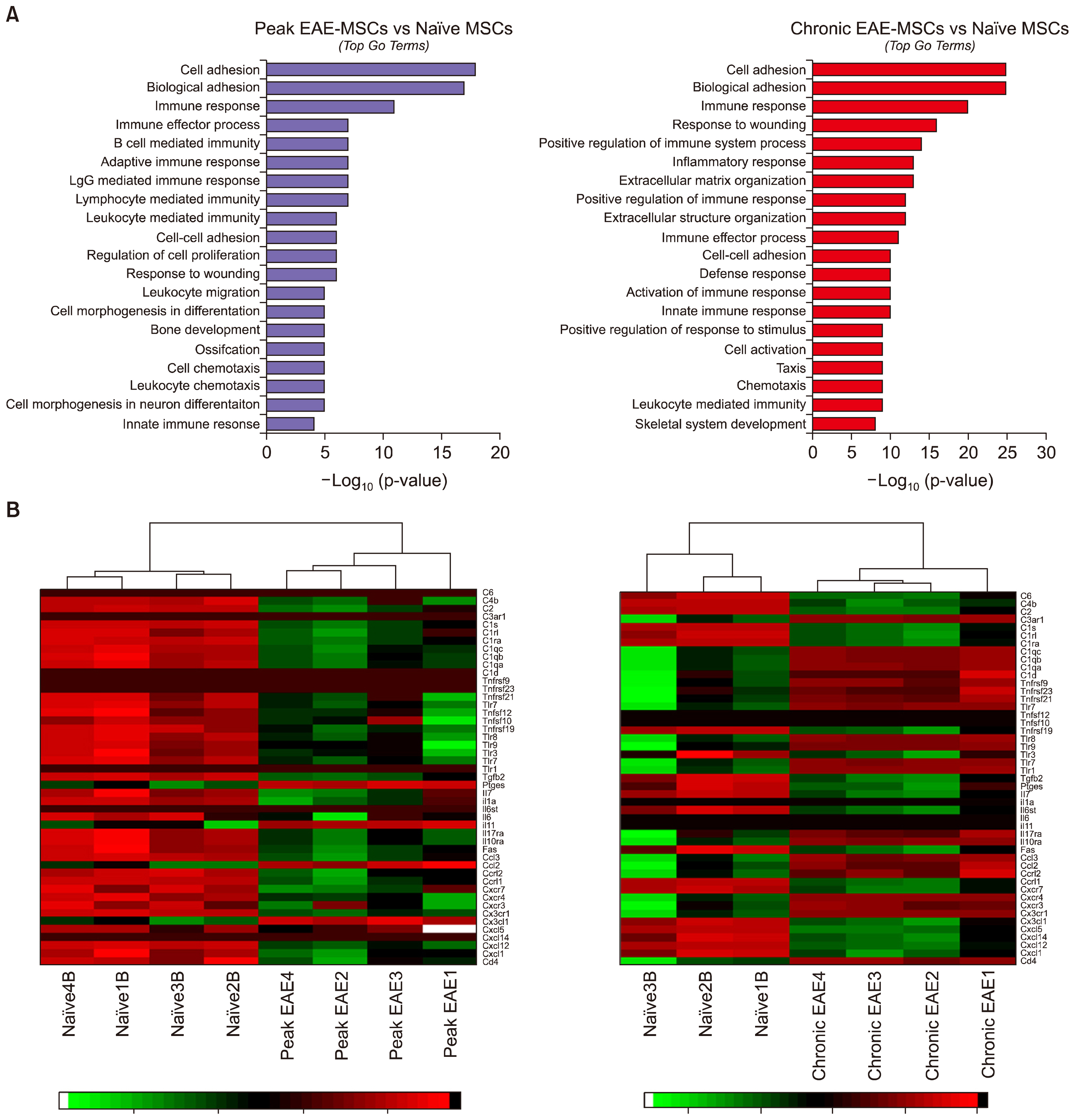
Fig. 3 EAE-MSCs show changes in genes associated with inflammation and immune activation. (A) Top 20 gene ontology terms (ranked by p-value) for DEGs found between peak EAE-MSCs and naïve MSCs (left) or chronic EAE-MSCs and naïve MSCs (right). Most GO terms relate to immune processes, particularly immune activation and immune response. (B) Heatmap showing immune genes differentially expressed between peak EAE-MSCs and naïve MSCs (left) or chronic EAE-MSCs and naïve MSCs (right). Many inflammatory genes, including chemokines, cytokines and elements of the complement system, are up regulated in EAE-MSCs.

Fig. 4 EAE-MSCs differentially regulate neural cell development and oligodendrocyte formation. (A) Top pathways (ranked by p-value) associated with the DEGs found between peak EAE-MSCs and naïve MSCs (left) or chronic EAE-MSCs and naïve MSCs (right). The number in parenthesis next to the pathway identifies statistical ranking assigned by MetaCore. Notable immune and neural cell pathways have been highlighted in green and red respectively. (B) Heatmap showing expression changes between peak EAE-MSCs and naïve MSCs (left) or chronic EAE-MSCs and naïve MSCs (right) for genes important in regulating neural cell development. (C) Neural cell cultures treated with conditioned medium (CM) from naïve MSCs had significantly higher number of MBP+ oligodendrocytes compared to control cultures treated with unconditioned medium, whereas cultures treated with EAE-MSC CM had a higher number of GFAP+ astrocytes relative to controls and they appear more reactive (insert). Scale bar=30 μm, data shown in graph=mean+SEM, **p<0.01, One-way ANOVA.

Fig. 5 Changes in gene expression in BM-MSCs throughout EAE. (A) Volcano plot depicting differentially expressed genes (DEGs) measured by RNA-Seq analysis of peak EAE-MSCs versus chronic EAE-MSCs. (B) Top 10 gene ontology (GO) terms ranked by p-value associated with the DEGs found between peak and chronic EAE-MSCs. (C) Heatmap showing immune genes differentially expressed between peak EAE-MSCs and chronic EAE-MSCs. Many inflammatory genes are up regulated in chronic EAE-MSCs compared to peak EAE-MSCs. (D) Venn diagram illustrating similarities in DEGs found between naïve, peak, and chronic EAE-MSCs. (E) Top 10 transcriptional regulators (identified through MetaCore) associated with the DEGs found between peak EAE-MSCs and naïve MSCs (left) or chronic EAE-MSCs and naïve MSCs (right). Transcription factors (TF) are ranked according to their respective z-score (the level of connectivity of the TF to the DEG list), with the number of DEGs associated with that TF indicated above each bar.
Cited by 2 articles
-
Mesenchymal Stem Cell-Derived Exosomes: A Promising Therapeutic Ace Card to Address Autoimmune Diseases
Hussein Baharlooi, Maryam Azimi, Zahra Salehi, Maryam Izad
Int J Stem Cells. 2019;13(1):13-23. doi: 10.15283/ijsc19108.Effect of Substrate Topography and Chemistry on Human Mesenchymal Stem Cell Markers: A Transcriptome Study
Bo Zhang, Naresh Kasoju, Qiongfang Li, Jinmin Ma, Aidong Yang, Zhanfeng Cui, Hui Wang, Hua Ye
Int J Stem Cells. 2019;12(1):84-94. doi: 10.15283/ijsc18102.
Reference
-
References
1. Uccelli A, Moretta L, Pistoia V. Mesenchymal stem cells in health and disease. Nat Rev Immunol. 2008; 8:726–736. DOI: 10.1038/nri2395.
Article2. Hass R, Kasper C, Böhm S, Jacobs R. Different populations and sources of human mesenchymal stem cells (MSC): A comparison of adult and neonatal tissue-derived MSC. Cell Commun Signal. 2011; 9:12. DOI: 10.1186/1478-811X-9-12. PMID: 21569606. PMCID: 3117820.
Article3. Kassis I, Vaknin-Dembinsky A, Karussis D. Bone marrow mesenchymal stem cells: agents of immunomodulation and neuroprotection. Curr Stem Cell Res Ther. 2011; 6:63–68. DOI: 10.2174/157488811794480762.
Article4. Sargent A, Miller RH. MSC Therapeutics in chronic inflammation. Curr Stem Cell Rep. 2016; 2:168–173. DOI: 10.1007/s40778-016-0044-6.
Article5. Stagg J, Galipeau J. Mechanisms of immune modulation by mesenchymal stromal cells and clinical translation. Curr Mol Med. 2013; 13:856–867. DOI: 10.2174/1566524011313050016. PMID: 23642066.
Article6. De Miguel MP, Fuentes-Julián S, Blázquez-Martínez A, Pascual CY, Aller MA, Arias J, Arnalich-Montiel F. Immunosuppressive properties of mesenchymal stem cells: advances and applications. Curr Mol Med. 2012; 12:574–591. DOI: 10.2174/156652412800619950. PMID: 22515979.
Article7. Rivera FJ, Couillard-Despres S, Pedre X, Ploetz S, Caioni M, Lois C, Bogdahn U, Aigner L. Mesenchymal stem cells instruct oligodendrogenic fate decision on adult neural stem cells. Stem Cells. 2006; 24:2209–2219. DOI: 10.1634/stemcells.2005-0614. PMID: 16763198.
Article8. Bai L, Lennon DP, Eaton V, Maier K, Caplan AI, Miller SD, Miller RH. Human bone marrow-derived mesenchymal stem cells induce Th2-polarized immune response and promote endogenous repair in animal models of multiple sclerosis. Glia. 2009; 57:1192–1203. DOI: 10.1002/glia.20841. PMID: 19191336. PMCID: 2706928.
Article9. Jadasz JJ, Kremer D, Göttle P, Tzekova N, Domke J, Rivera FJ, Adjaye J, Hartung HP, Aigner L, Küry P. Mesenchymal stem cell conditioning promotes rat oligodendroglial cell maturation. PLoS One. 2013; 8:e71814. DOI: 10.1371/journal.pone.0071814. PMID: 23951248. PMCID: 3741203.
Article10. Bai L, Lennon DP, Caplan AI, DeChant A, Hecker J, Kranso J, Zaremba A, Miller RH. Hepatocyte growth factor mediates mesenchymal stem cell–induced recovery in multiple sclerosis models. Nat Neurosci. 2012; 15:862–870. DOI: 10.1038/nn.3109. PMID: 22610068. PMCID: 3427471.
Article11. Miller RH, Bai L, Lennon DP, Caplan AI. The potential of mesenchymal stem cells for neural repair. Discov Med. 2010; 9:236–242. PMID: 20350491.12. Gold R, Linington C, Lassmann H. Understanding pathogenesis and therapy of multiple sclerosis via animal models: 70 years of merits and culprits in experimental autoimmune encephalomyelitis research. Brain. 2006; 129:1953–1971. DOI: 10.1093/brain/awl075.
Article13. Zappia E, Casazza S, Pedemonte E, Benvenuto F, Bonanni I, Gerdoni E, Giunti D, Ceravolo A, Cazzanti F, Frassoni F, Mancardi G, Uccelli A. Mesenchymal stem cells ameliorate experimental autoimmune encephalomyelitis inducing T-cell anergy. Blood. 2005; 106:1755–1761. DOI: 10.1182/blood-2005-04-1496. PMID: 15905186.
Article14. Zhang J, Li Y, Chen J, Cui Y, Lu M, Elias SB, Mitchell JB, Hammill L, Vanguri P, Chopp M. Human bone marrow stromal cell treatment improves neurological functional recovery in EAE mice. Exp Neurol. 2005; 195:16–26. DOI: 10.1016/j.expneurol.2005.03.018. PMID: 15904921.
Article15. Zhang J, Li Y, Lu M, Cui Y, Chen J, Noffsinger L, Elias SB, Chopp M. Bone marrow stromal cells reduce axonal loss in experimental autoimmune encephalomyelitis mice. J Neurosci Res. 2006; 84:587–595. DOI: 10.1002/jnr.20962. PMID: 16773650.
Article16. Al Jumah MA, Abumaree MH. The immunomodulatory and neuroprotective effects of mesenchymal stem cells (MSCs) in experimental autoimmune encephalomyelitis (EAE): a model of multiple sclerosis (MS). Int J Mol Sci. 2012; 13:9298–9331. DOI: 10.3390/ijms13079298. PMID: 22942767. PMCID: 3430298.
Article17. Meamar R, Nematollahi S, Dehghani L, Mirmosayyeb O, Shayegannejad V, Basiri K, Tanhaei AP. The role of stem cell therapy in multiple sclerosis: An overview of the current status of the clinical studies. Adv Biomed Res. 2016; 5:46. DOI: 10.4103/2277-9175.178791. PMID: 27110543. PMCID: 4817403.
Article18. Sargent A, Bai L, Shano G, Karl M, Garrison E, Ranasinghe L, Planchon SM, Cohen J, Miller RH. CNS disease diminishes the therapeutic functionality of bone marrow mesenchymal stem cells. Exp Neurol. 2017; 295:222–232. DOI: 10.1016/j.expneurol.2017.06.013. PMID: 28602834. PMCID: 5536847.
Article19. Korn T, Mitsdoerffer M, Croxford AL, Awasthi A, Dardalhon VA, Galileos G, Vollmar P, Stritesky GL, Kaplan MH, Waisman A, Kuchroo VK, Oukka M. IL-6 controls Th17 immunity in vivo by inhibiting the conversion of conventional T cells into Foxp3+ regulatory T cells. Proc Natl Acad Sci U S A. 2008; 105:18460–18465. DOI: 10.1073/pnas.0809850105. PMID: 19015529. PMCID: 2587589.
Article20. Li Y, Lin F. Mesenchymal stem cells are injured by complement after their contact with serum. Blood. 2012; 120:3436–3443. DOI: 10.1182/blood-2012-03-420612. PMID: 22966167. PMCID: 3482856.
Article21. Waterman RS, Tomchuck SL, Henkle SL, Betancourt AM. A new mesenchymal stem cell (MSC) paradigm: polarization into a pro-inflammatory MSC1 or an Immunosuppressive MSC2 phenotype. PLoS One. 2010; 5:e10088. DOI: 10.1371/journal.pone.0010088. PMID: 20436665. PMCID: 2859930.
Article22. Wen S, Li H, Liu J. Dynamic signaling for neural stem cell fate determination. Cell Adh Migr. 2009; 3:107–117. DOI: 10.4161/cam.3.1.7602. PMID: 19262166. PMCID: 2675157.
Article23. Grinspan JB, Edell E, Carpio DF, Beesley JS, Lavy L, Pleasure D, Golden JA. Stage-specific effects of bone morphogenetic proteins on the oligodendrocyte lineage. J Neurobiol. 2000; 43:1–17. DOI: 10.1002/(SICI)1097-4695(200004)43:1<1::AID-NEU1>3.0.CO;2-0. PMID: 10756062.
Article24. Stipursky J, Gomes FC. TGF-beta1/SMAD signaling induces astrocyte fate commitment in vitro: implications for radial glia development. Glia. 2007; 55:1023–1033. DOI: 10.1002/glia.20522. PMID: 17549683.
Article25. Palazuelos J, Klingener M, Aguirre A. TGFβ signaling regulates the timing of CNS myelination by modulating oligodendrocyte progenitor cell cycle exit through SMAD3/4/FoxO1/Sp1. J Neurosci. 2014; 34:7917–7930. DOI: 10.1523/JNEUROSCI.0363-14.2014. PMID: 24899714. PMCID: 4044250.
Article26. Hsieh J, Aimone JB, Kaspar BK, Kuwabara T, Nakashima K, Gage FH. IGF-I instructs multipotent adult neural progenitor cells to become oligodendrocytes. J Cell Biol. 2004; 164:111–122. DOI: 10.1083/jcb.200308101. PMID: 14709544. PMCID: 2171962.
Article27. Auletta JJ, Bartholomew AM, Maziarz RT, Deans RJ, Miller RH, Lazarus HM, Cohen JA. The potential of mesenchymal stromal cells as a novel cellular therapy for multiple sclerosis. Immunotherapy. 2012; 4:529–547. DOI: 10.2217/imt.12.41. PMID: 22642335. PMCID: 3381871.
Article28. Bonab MM, Sahraian MA, Aghsaie A, Karvigh SA, Hosseinian SM, Nikbin B, Lotfi J, Khorramnia S, Motamed MR, Togha M, Harirchian MH, Moghadam NB, Alikhani K, Yadegari S, Jafarian S, Gheini MR. Autologous mesenchymal stem cell therapy in progressive multiple sclerosis: an open label study. Curr Stem Cell Res Ther. 2012; 7:407–414. DOI: 10.2174/157488812804484648. PMID: 23061813.
Article29. Yamout B, Hourani R, Salti H, Barada W, El-Hajj T, Al-Kutoubi A, Herlopian A, Baz EK, Mahfouz R, Khalil-Hamdan R, Kreidieh NM, El-Sabban M, Bazarbachi A. Bone marrow mesenchymal stem cell transplantation in patients with multiple sclerosis: a pilot study. J Neuroimmunol. 2010; 227:185–189. DOI: 10.1016/j.jneuroim.2010.07.013. PMID: 20728948.
Article30. Connick P, Kolappan M, Crawley C, Webber DJ, Patani R, Michell AW, Du MQ, Luan SL, Altmann DR, Thompson AJ, Compston A, Scott MA, Miller DH, Chandran S. Autologous mesenchymal stem cells for the treatment of secondary progressive multiple sclerosis: an open-label phase 2a proof-of-concept study. Lancet Neurol. 2012; 11:150–156. DOI: 10.1016/S1474-4422(11)70305-2. PMID: 22236384. PMCID: 3279697.
Article31. Karussis D, Karageorgiou C, Vaknin-Dembinsky A, Gowda-Kurkalli B, Gomori JM, Kassis I, Bulte JW, Petrou P, Ben-Hur T, Abramsky O, Slavin S. Safety and immunological effects of mesenchymal stem cell transplantation in patients with multiple sclerosis and amyotrophic lateral sclerosis. Arch Neurol. 2010; 67:1187–1194. DOI: 10.1001/archneurol.2010.248. PMID: 20937945. PMCID: 3036569.
Article32. Dulamea A. Mesenchymal stem cells in multiple sclerosis - translation to clinical trials. J Med Life. 2015; 8:24–27. PMID: 25914733. PMCID: 4397514.33. Mallam E, Kemp K, Wilkins A, Rice C, Scolding N. Characterization of in vitro expanded bone marrow-derived mesenchymal stem cells from patients with multiple sclerosis. Mult Scler. 2010; 16:909–918. DOI: 10.1177/1352458510371959. PMID: 20542920.
Article34. Mazzanti B, Aldinucci A, Biagioli T, Barilaro A, Urbani S, Dal Pozzo S, Amato MP, Siracusa G, Crescioli C, Manuelli C, Bosi A, Saccardi R, Massacesi L, Ballerini C. Differences in mesenchymal stem cell cytokine profiles between MS patients and healthy donors: implication for assessment of disease activity and treatment. J Neuroimmunol. 2008; 199:142–150. DOI: 10.1016/j.jneuroim.2008.05.006. PMID: 18562015.
Article35. Redondo J, Sarkar P, Kemp K, Virgo PF, Pawade J, Norton A, Emery DC, Guttridge MG, Marks DI, Wilkins A, Scolding NJ, Rice CM. Reduced cellularity of bone marrow in multiple sclerosis with decreased MSC expansion potential and premature ageing in vitro. Mult Scler. 2017; 1352458517711276. DOI: 10.1177/1352458517711276. PMID: 28548004.
Article36. de Oliveira GL, de Lima KW, Colombini AM, Pinheiro DG, Panepucci RA, Palma PV, Brum DG, Covas DT, Simões BP, de Oliveira MC, Donadi EA, Malmegrim KC. Bone marrow mesenchymal stromal cells isolated from multiple sclerosis patients have distinct gene expression profile and decreased suppressive function compared with healthy counterparts. Cell Transplant. 2015; 24:151–165. DOI: 10.3727/096368913X675142.
Article37. Siegel G, Kluba T, Hermanutz-Klein U, Bieback K, Northoff H, Schäfer R. Phenotype, donor age and gender affect function of human bone marrow-derived mesenchymal stromal cells. BMC Med. 2013; 11:146. DOI: 10.1186/1741-7015-11-146. PMID: 23758701. PMCID: 3694028.
Article38. Cohen JA. Mesenchymal stem cell transplantation in multiple sclerosis. J Neurol Sci. 2013; 333:43–49. DOI: 10.1016/j.jns.2012.12.009. PMID: 23294498. PMCID: 3624046.
Article39. Miller RH. Regulation of oligodendrocyte development in the vertebrate CNS. Prog Neurobiol. 2002; 67:451–467. DOI: 10.1016/S0301-0082(02)00058-8. PMID: 12385864.
Article
- Full Text Links
-
- Actions
-
Cited
- CITED
-
- Close
- Share
-
- Similar articles
-
- Role of Mesenchymal Stem Cells in Patients with Nontraumatic Osteonecrosis of the Femoral Head
- Insights into the signal transduction pathways of mouse lung type II cells revealed by transcription factor profiling in the transcriptome
- Recent Trends and Strategies in Stem Cell Therapy for Alzheimer's Disease
- Stem Cells and Niemann Pick Disease
- Nineteen Types of WNT Genes Mediate The Differentiation of C3H10T1/2 Cell Lines
