J Adv Prosthodont.
2018 Jun;10(3):218-226. 10.4047/jap.2018.10.3.218.
Color comparison between non-vital and vital teeth
- Affiliations
-
- 1Department of Prosthetic Dentistry and Dental Materials, Division of Dental Propaedeutics and Esthetic Dentistry, Faculty of Dentistry, Iuliu Haţieganu University of Medicine and Pharmacy, Cluj-Napoca, Romania.
- 2Department of Medical Education, Division of Medical Informatics and Biostatistics, Faculty of Medicine, Iuliu Haţieganu University of Medicine and Pharmacy, Cluj-Napoca, Romania. hcolosi@umfcluj.ro
- KMID: 2413491
- DOI: http://doi.org/10.4047/jap.2018.10.3.218
Abstract
- PURPOSE
The aim of this study was to define a color space of non-vital teeth and to compare it with the color space of matched vital teeth, recorded in the same patients.
MATERIALS AND METHODS
In a group of 218 patients, with the age range from 17 to 70, the middle third of the buccal surface of 359 devitalized teeth was measured using a clinical spectrophotometer (Vita Easyshade Advance). Lightness (L*), chromatic parameters (a*, b*), chroma (C*), hue angle (h) and the closest Vita shade in Classical and 3D Master codifications were recorded. For each patient, the same data were recorded in a vital reference tooth. The measurements were performed by the same operator with the same spectrophotometer, using a standardized protocol for color evaluation.
RESULTS
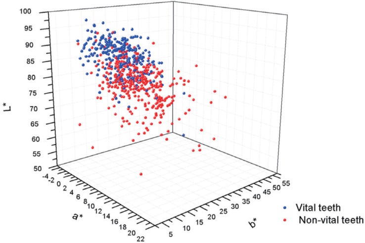
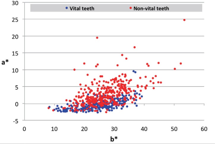
The color coordinates of non-vital teeth varied as follows: lightness L*: 52.83-92.93, C*: 8.23-58.90, h: 51.20-101.53, a*: −2.53-24.80, b*: 8.10-53.43. For the reference vital teeth, the ranges of color parameters were: L*: 60.90-97.16, C*: 8.43-39.23, h: 75.30-101.13, a*: −2.36-9.60, b*: 8.36-39.23. The color differences between vital and non-vital teeth depended on tooth group, but not on patient age.
CONCLUSION
Non-vital teeth had a wider color space than vital ones. Non-vital teeth were darker (decreased lightness), more saturated (increased chroma), and with an increased range of the hue interval. An increased tendency towards positive values on the a* and b* axes suggested redder and yellower non-vital teeth compared to vital ones.
MeSH Terms
Figure
Reference
-
1. Chu SJ, Trushkowsky RD, Paravina RD. Dental color matching instruments and systems. Review of clinical and research aspects. J Dent. 2010; 38:e2–e16. PMID: 20621154.
Article2. Tour Savadkouhi S, Fazlyab M. Discoloration potential of endodontic sealers: A brief review. Iran Endod J. 2016; 11:250–254. PMID: 27790251.3. Ioannidis K, Beltes P, Lambrianidis T, Kapagiannidis D, Karagiannis V. Crown discoloration induced by endodontic sealers: spectrophotometric measurement of Commission International de I'Eclairage's L*, a*, b* chromatic parameters. Oper Dent. 2013; 38:E1–E12.4. Mayekar SM. Shades of a color. Illusion or reality? Dent Clin North Am. 2001; 45:155–172. viiPMID: 11210693.5. Rimmer SE, Mellor AC. Patients' perceptions of esthetics and technical quality in crowns and fixed partial dentures. Quintessence Int. 1996; 27:155–162. PMID: 9063227.6. Commission Internationale de l'Eclairage. CIE Technical Report: Colorimetry. Available from: https://www.cdvplus.cz/file/3-publikace-cie15-2004/.7. Manauta J, Salat A. Out. In : Manauta J, Salat A, editors. Layers: An atlas of composite resin stratification. Milan: Quintessenza Edizioni;2012. Available from: http://www.dentalbooks.bg/PDF/Layers%20Stratification%20MANAUTA.pdf.8. Rubiño M, Garcia JA, Jiménez del, Romero J. Colour measurement of human teeth and evaluation of a colour guide. Color Res Appl. 1994; 19:19–22.9. Zhu H, Lei Y, Liao N. Color measurements of 1,944 anterior teeth of people in southwest of China-discreption. Zhonghua Kou Qiang Yi Xue Za Zhi. 2001; 36:285–288. PMID: 11718012.10. Joiner A, Hopkinson I, Deng Y, Westland S. A review of tooth colour and whiteness. J Dent. 2008; 36:S2–S7. PMID: 18646363.
Article11. Gerlach RW, Barker ML, Sagel PA. Objective and subjective whitening response of two self-directed bleaching systems. Am J Dent. 2002; 15:7A–12A.12. Xu Y. Research on the designing method of a special shade guide for tooth whitening. Hua Xi Kou Qiang Yi Xue Za Zhi. 2015; 33:478–483. PMID: 26688939.13. Gehrke P, Riekeberg U, Fackler O, Dhom G. Comparison of in vivo visual, spectrophotometric and colorimetric shade determination of teeth and implant-supported crowns. Int J Comput Dent. 2009; 12:247–263. PMID: 19715149.14. Da Silva JD, Park SE, Weber HP, Ishikawa-Nagai S. Clinical performance of a newly developed spectrophotometric system on tooth color reproduction. J Prosthet Dent. 2008; 99:361–368. PMID: 18456047.
Article15. Gómez-Polo C, Gómez-Polo M, Martínez Vázquez de Parga JA, Celemín Viñuela A. Study of the most frequent natural tooth colors in the Spanish population using spectrophotometry. J Adv Prosthodont. 2015; 7:413–422. PMID: 26816571.
Article16. Yuan JC, Brewer JD, Monaco EA Jr, Davis EL. Defining a natural tooth color space based on a 3-dimensional shade system. J Prosthet Dent. 2007; 98:110–119. PMID: 17692592.
Article17. O'Brien WJ, Hemmendinger H, Boenke KM, Linger JB, Groh CL. Color distribution of three regions of extracted human teeth. Dent Mater. 1997; 13:179–185. PMID: 9758972.18. Hasegawa A, Ikeda I, Kawaguchi S. Color and translucency of in vivo natural central incisors. J Prosthet Dent. 2000; 83:418–423. PMID: 10756291.
Article19. Ahmad Rana N, Farid H, Saad Shinwari M, Anwar A. Identification of tooth shade in various age groups of Pakistani population using Vita Easyshade. J Khyber Coll Dent. 2014; 5:25–28.20. Elamin HO, Abubakr NH, Ibrahim YE. Identifying the tooth shade in group of patients using Vita Easyshade. Eur J Dent. 2015; 9:213–217. PMID: 26038652.
Article21. Rodrigues S, Shetty SR, Prithviraj DR. An evaluation of shade differences between natural anterior teeth in different age groups and gender using commercially available shade guides. J Indian Prosthodont Soc. 2012; 12:222–230. PMID: 24293919.
Article22. Pop-Ciutrila IS, Ghinea R, Perez Gomez MDM, Colosi HA, Dudea D, Badea M. Dentine scattering, absorption, transmittance and light reflectivity in human incisors, canines and molars. J Dent. 2015; 43:1116–1124. PMID: 26149064.
Article23. Goodkind RJ, Schwabacher WB. Use of a fiber-optic colorimeter for in vivo color measurements of 2830 anterior teeth. J Prosthet Dent. 1987; 58:535–542. PMID: 3479551.
Article24. Ghinea R, Pérez MM, Herrera LJ, Rivas MJ, Yebra A, Paravina RD. Color difference thresholds in dental ceramics. J Dent. 2010; 38:e57–e64. PMID: 20670670.
Article25. Paravina RD, Ghinea R, Herrera LJ, Bona AD, Igiel C, Linninger M, Sakai M, Takahashi H, Tashkandi E, Perez Mdel M. Color difference thresholds in dentistry. J Esthet Restor Dent. 2015; 27:S1–S9. PMID: 25886208.
Article26. Gómez-Polo C, Gómez-Polo M, Celemin-Viñuela A, Martínez Vázquez De Parga JA. Differences between the human eye and the spectrophotometer in the shade matching of tooth colour. J Dent. 2014; 42:742–745. PMID: 24140995.
Article27. Olms C, Setz JM. The repeatability of digital shade measurement-a clinical study. Clin Oral Investig. 2013; 17:1161–1166.
Article28. Klaff D. Blending incremental and stratified layering techniques to produce an esthetic posterior composite resin restoration with a predictable prognosis. J Esthet Restor Dent. 2001; 13:101–113. PMID: 11499445.
Article29. Odioso LL, Gibb RD, Gerlach RW. Impact of demographic, behavioral, and dental care utilization parameters on tooth color and personal satisfaction. Compend Contin Educ Dent Suppl. 2000; S35–S41. PMID: 11908408.
- Full Text Links
- Actions
-
Cited
- CITED
-
- Close
- Share
- Similar articles
-
- Effect of Kegel Exercise on Vital Capacity According to the Position: A Preliminary Study
- Prediction of Normal Values of Vital Capacity in Teenagers
- Color Change in Tooth Induced by Various Calcium Silicate-Based Pulp-Capping Materials
- Treatment of non-vital immature teeth with amoxicillin-containing triple antibiotic paste resulting in apexification
- Color Comparison of Maxillary Primary Anterior Teeth and Various Composite Resins using a Spectrophotometer


