Arch Hand Microsurg.
2018 Jun;23(2):121-125. 10.12790/ahm.2018.23.2.121.
An Iatrogenic Flap Necrosis Case: Factors to Consider for Successful Flap Elevation
- Affiliations
-
- 1Department of Plastic and Reconstructive Surgery, Ajou University School of Medicine, Suwon, Korea. i00325@live.co.kr
- KMID: 2412482
- DOI: http://doi.org/10.12790/ahm.2018.23.2.121
Abstract
- The free flap technique has been used for lower limb reconstruction. Still, the free flap itself and related surgical procedures are challenging process, because there's a lot of factors to consider for successful outcome. We report a case of total flap necrosis due to inadequate flap elevation on the left ankle of a young man. Although the shape of the elevated flap was not fit into rectangular shape exactly, the length to width ratio of the flap was almost 2.25:1. The circulation to the tissue was not enough to flap survival, due to inadequate incision. To prevent flap mismanagement, all the surgical procedure should be performed based on a thorough understanding of flap related knowledge, such as length-width ratio restriction of elevated flap, angiosome and proper surgical incision. Also we recommend swell-timed consultation with surgeons trained in microsurgery for guidance regarding flap evaluation and its subsequent management.
Keyword
Figure
-
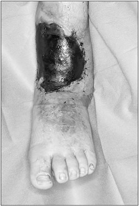
Fig. 1 Total flap necrosis of a thoracodorsal artery perforator free flap at 1 year after surgery. Necrosis was due to inadequate U-shaped flap elevation.
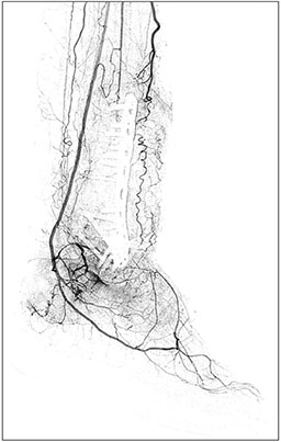
Fig. 2 Angiography of the patient's lower extremity. The anterior tibial artery supplies the flap. The posterior tibial artery is intact. Well-developed collateral arteries are present.
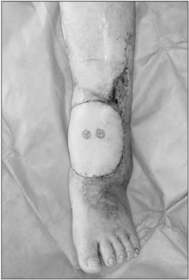
Fig. 3 Postoperative clinical photograph after removing all stitches.
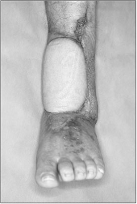
Fig. 4 Photograph of the patient at 3.5 months after surgery reveals no complications.
Reference
-
1. Bradshaw K, Wagels M. Perfusion of muscle flaps independent of the anatomical vascular pedicle: pedicle autonomy. J Plast Reconstr Aesthet Surg. 2017; 70:1547–1555.
Article2. Jordan DJ, Malahias M, Hindocha S, Juma A. Flap decisions and options in soft tissue coverage of the lower limb. Open Orthop J. 2014; 8:Suppl 2: M5. 423–432.
Article3. Attinger CE, Evans KK, Bulan E, Blume P, Cooper P. Angiosomes of the foot and ankle and clinical implications for limb salvage: reconstruction, incisions, and revascularization. Plast Reconstr Surg. 2006; 117:7 Suppl. 261S–293S.
Article4. Attinger C, Cooper P, Blume P, Bulan E. The safest surgical incisions and amputations applying the angiosome principles and using the doppler to assess the arterial-arterial connections of the foot and ankle. Foot Ankle Clin. 2001; 6:745–799.
Article
- Full Text Links
-
- Actions
-
Cited
- CITED
-
- Close
- Share
-
- Similar articles
-
- Flap thinning: Defatting after conventional elevation
- Safety of Elevation from Superficial Fascial Plane versus Traditional Deep Fascial Plane for Flap Elevation in a Porcine Model
- Successful reconstruction using a de-epithelialized rectangular flap on a nipple necrosis site after DIEP flap-based breast reconstruction: a case report
- Effect of prefabrication on the survival of venous island flap
- Perineum-based Pediculated Scrotal Flap for Reconstructive Urethral Surgery
