Evaluation of the ureteral jet in dogs by using color Doppler ultrasonography
- Affiliations
-
- 1Department of Veterinary Medical Imaging, College of Veterinary Medicine, Seoul National University, Seoul 08826, Korea. mcchoi@snu.ac.kr
- 2Ilsan Animal Medical Center, Goyang 10368, Korea.
- KMID: 2412458
- DOI: http://doi.org/10.4142/jvs.2017.18.3.399
Abstract
- Ureteral jets are the result of a forceful ejection of urine from the vesicoureteral junction into the urinary bladder. By using color Doppler ultrasonography (US), we aimed to identify distinct ureteral jets in dogs, provide insight into ureteral obstruction, and facilitate study of urodynamics and vesicoureteric sphincter function via pulsed Doppler US. Color Doppler US was applied to detect urinary flow from the right ureteral orifices in eight healthy beagles. Under anesthesia, 0.9% saline (2.5 mL/kg/h) and furosemide (0.5 mg/kg) were administered intravenously to assist in detection of distinct ureteral jets and examine their frequency, velocity, duration, and waveform. In all dogs, ureteral jets were visualized under diuresis and anesthesia within 2 to 5 min (mean 3.57 ± 0.90 min) of the furosemide injection. Mean frequency, peak velocity, and duration of right ureteral jets in seven dogs in whom six ureteral jet waveform patterns were identified were 9.86 ± 3.09 jets/min, 34.07 ± 10.02 cm/sec, and 2.82 ± 1.08 sec, respectively. During the 10 min period starting 10 min after the initial jet appeared, only three waveforms were identified. Color Doppler US of ureteral jets may aid in assessing vesicoureteric sphincter function and ureteral abnormalities, such as ureteral obstruction, in dogs.
MeSH Terms
Figure
-
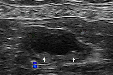
Fig. 1 Transverse ultrasonographic image of the urinary bladder showing two small elevations of the mucosal surface representing the ureteral orifices (arrows) on the dorsal aspect.
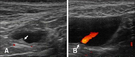
Fig. 2 Oblique ultrasonographic image of the urinary bladder clearly showing the orifice (A) and the jet flow (B). The anechoic area within the elevation represents the right ureter (arrows).
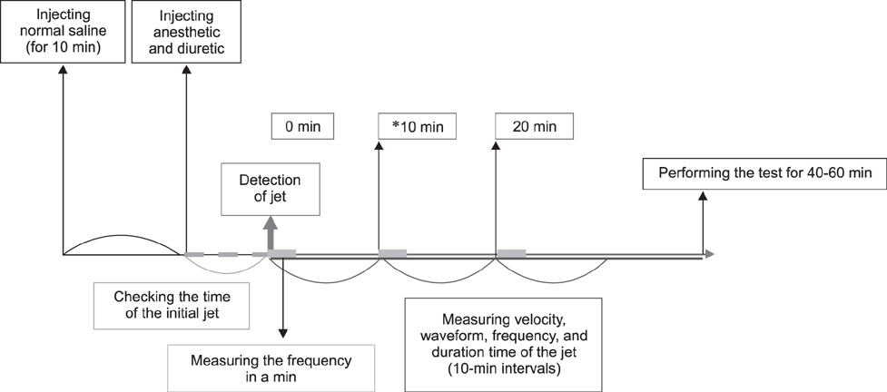
Fig. 3 Schematic diagram of the experimental procedure. *Period in which the jet is identified most distinctively.
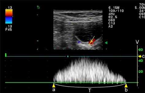
Fig. 4 Spectral Doppler image of the right ureteral orifice. The peak velocity (c) is approximately 47.8 cm/sec, and the duration time (T) is 2.0 sec. The waveform is monophasic. a, point of commencement of the Doppler signal; b, termination of signal; V, velocity.
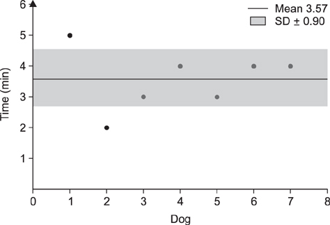
Fig. 5 The mean and standard deviation of the initial time of appearance of ureteral jets. Note that the jet phenomenon occurred in each dog (except Dog 8) within 5 min of injection of the diuretic.
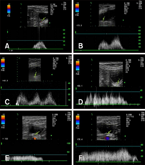
Fig. 6 Six distinctive waveform patterns of the ureteral jet. (A) Monophasic waveform. (B) Biphasic waveform. (C) Triphasic waveform. (D) Polyphasic waveform. (E) Square waveform. (F) Continuous waveform.
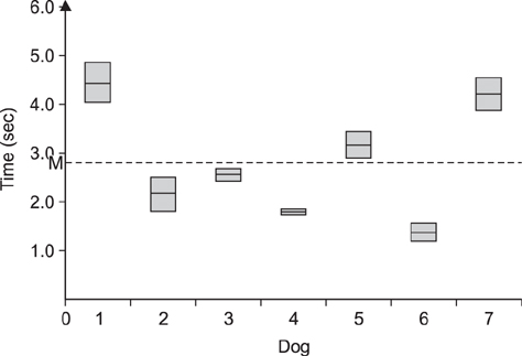
Fig. 7 Mean ureteral jet duration in each dog and mean duration in all dogs (M).
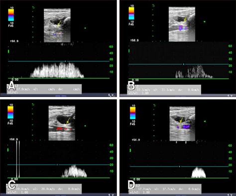
Fig. 8 Spectral Doppler images of changing jet appearance and duration.
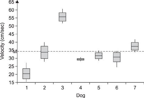
Fig. 9 Mean ureteral jet peak velocity in each dog and mean value in all dogs (M).
Reference
-
1. Akçar N, Özkan IR, Adapınar B, Kaya T. Doppler sonography in the diagnosis of urinary tract obstruction by stone. J Clin Ultrasound. 2004; 32:286–293.
Article2. Baker SM, Middleton WD. Color Doppler sonography of ureteral jets in normal volunteers: importance of the relative specific gravity of urine in the ureter and bladder. AJR Am J Roentgenol. 1992; 159:773–775.
Article3. Burge HJ, Middleton WD, McClennan BL, Hildebolt CF. Ureteral jets in healthy subjects and in patients with unilateral ureteral calculi: comparison with color Doppler US. Radiology. 1991; 180:437–442.
Article4. Catalano O, De Sena G, Nunziata A. [The color Doppler US evaluation of the ureteral jet in patients with urinary colic]. Radiol Med. 1998; 95:614–617. Italian.5. Cox IH, Erickson SJ, Foley WD, Dewire DM. Ureteric jets: evaluation of normal flow dynamics with color Doppler sonography. AJR Am J Roentgenol. 1992; 158:1051–1055.
Article6. Curry NS, Gobien RP, Schabel SI. Minimal-dilatation obstructive nephropathy. Radiology. 1982; 143:531–534.
Article7. Daniel EE, Tomita T, Tsuchida S, Watanabe M. Sphincters: Normal Function-Changes in Diseases. Tokyo: CRC Press;1992. p. 295–304.8. Delair SM, Kurzrock EA. Clinical utility of ureteral jets: disparate opinions. J Endourol. 2006; 20:111–114.
Article9. Deyoe LA, Cronan JJ, Breslaw BH, Ridlen MS. New techniques of ultrasound and color Doppler in the prospective evaluation of acute renal obstruction. Do they replace the intravenous urogram. Abdom Imaging. 1995; 20:58–63.
Article10. Dubbins PA, Kurtz AB, Darby J, Goldberg BB. Ureteric jet effect: the echographic appearance of urine entering the bladder. A means of identifying the bladder trigone and assessing ureteral function. Radiology. 1981; 140:513–515.
Article11. Elejalde BR, de Elejalde MM. Ureteral ejaculation of urine visualized by ultrasound. J Clin Ultrasound. 1983; 11:475–476.
Article12. Erwin BC, Carroll BA, Sommer FG. Renal colic: the role of ultrasound in initial evaluation. Radiology. 1984; 152:147–150.
Article13. Grisi G, Stacul F, Cuttin R, Rimondini A, Meduri S, Dalla Palma L. Cost analysis of different protocols for imaging a patient with acute flank pain. Eur Radiol. 2000; 10:1620–1627.
Article14. Jafri SZ, Madrazo BL, Miller JH. Color Doppler ultrasound of the genitourinary tract. Curr Opin Radiol. 1992; 4:16–23.15. Jequier S, Paltiel H, Lafortune M. Ureterovesical jets in infants and children: duplex and color Doppler US studies. Radiology. 1990; 175:349–353.
Article16. Jubb KVF, Kennedy PC, Palmer N. Pathology of Domestic Animals. 3rd ed. Vol. 2. San Diego: Academic Press;1985. p. 390–392.17. Kamholtz RG, Cronan JJ, Dorfman GS. Obstruction and the minimally dilated renal collecting system: US evaluation. Radiology. 1989; 170:51–53.
Article18. Koelliker SL, Cronan JJ. Acute urinary tract obstruction: imaging update. Urol Clin North Am. 1997; 24:571–582.19. Kremer H, Dobrinski W, Mikyska M, Baumgärtner M, Zöllner N. Ultrasonic in vivo and in vitro studies on the nature of the ureteral jet phenomenon. Radiology. 1982; 142:175–177.
Article20. Kremkau FW, Gramiak R, Carstensen EL, Shah PM, Kramer DH. Ultrasonic detection of cavitation at catheter tips. Am J Roentgenol Radium Ther Nucl Med. 1970; 110:177–183.
Article21. Laing FC, Benson CB, DiSalvo DN, Brown DL, Frates MC, Loughlin KR. Distal ureteral calculi: detection with vaginal US. Radiology. 1994; 192:545–548.
Article22. Laing FC, Jeffrey RB Jr, Wing VW. Ultrasound versus excretory urography in evaluating acute flank pain. Radiology. 1985; 154:613–616.
Article23. Lamb CR, Gregory SP. Ultrasonography of the ureterovesicular junction in the dog: a preliminary report. Vet Rec. 1994; 134:36–38.
Article24. Leung VY, Chu WC, Yeung Ck, Metreweli C. Doppler waveforms of the ureteric jet: an overview and implications for the presence of a functional sphincter at the vesicoureteric junction. Pediatr Radiol. 2007; 37:417–425.
Article25. Leung VYF, Chu WCW. Functional anatomy of the vesicoureteric junction: implication on the management of VUR/ UTI. In : Nelius T, editor. Recent Advances in the Field of Urinary Tract Infections. Chapt. 6. InTech;2013.26. Leung VY, Metreweli C, Yeung CK. The ureteric jet Doppler waveform as an indicator of vesicoureteric sphincter function in adults and children. An observational study. Ultrasound Med Biol. 2002; 28:865–872.
Article27. Leung VY, Metreweli C, Yeung CK, Sihoe JD. Ureteric jet in the anaesthetised child. Ultrasound Med Biol. 2003; 29:1237–1240.
Article28. Marshall JL, Johnson ND, De Campo MP. Vesicoureteric reflux in children: prediction with color Doppler imaging. Work in progress. Radiology. 1990; 175:355–358.
Article29. Matsuda T, Saitoh M. Detection of the urine jet phenomenon using Doppler color flow mapping. Int J Urol. 1995; 2:232–234.
Article30. Meltzer RS, Tickner EG, Sahines TP, Popp RL. The source of ultrasound contrast effect. J Clin Ultrasound. 1980; 8:121–127.
Article31. Mostbeck GH, Zontsich T, Turetschek K. Ultrasound of the kidney: obstruction and medical diseases. Eur Radiol. 2001; 11:1878–1889.
Article32. Noordzij JW, Dabhoiwala NF. A view on the anatomy of the ureterovesical junction. Scand J Urol Nephrol. 1993; 27:371–380.
Article33. Patel U, Kellett MJ. Ureteric drainage and peristalsis after stenting studied using colour Doppler ultrasound. Br J Urol. 1996; 77:530–535.
Article34. Price CI, Adler RS, Rubin JM. Ultrasound detection of differences in density: explanation of the ureteric jet phenomenon and implications for new ultrasound applications. Invest Radiol. 1989; 24:876–883.
Article35. Saita H, Matsukawa M, Fukushima H, Ohyama C, Nagata Y. Ultrasound diagnosis of ureteral stones: its usefulness with subsequent excretory urography. J Urol. 1988; 140:28–31.
Article36. Strehlau J, Winkler P, de la Roche J. The uretero-vesical jet as a functional diagnostic tool in childhood hydronephrosis. Pediatr Nephrol. 1997; 11:460–467.
Article37. Sweet CS, Silbergleit R, Sanders WP. MRI demonstration of ureteral jet effect in a patient with a spinal ganglioneuroma. Pediatr Radiol. 1995; 25:574–575.
Article38. Tal Z, Jaffe H, Rosenak D, Nadjari M, Hornstein E. Ureteric jet examination by color Doppler ultrasound versus IVP for the assessment of ureteric patency following pelvic surgery- a pilot study. Eur J Obstet Gynecol Reprod Biol. 1994; 54:119–122.
Article39. Timor-Tritsch IE, Haratz-Rubinstein N, Monteagudo A, Lerner JP, Murphy KE. Transvaginal color Doppler sonography of the ureteral jets: a method to detect ureteral patency. Obstet Gynecol. 1997; 89:113–117.
Article40. Van Arsdalen KN, Banner MP, Pollack HM. Radiographic imaging and urologic decision making in the management of renal and ureteral calculi. Urol Clin North Am. 1990; 17:171–190.
Article41. Wu CC, Yao WJ, Lin F Jr, Hsieh HL, Hwang MH. Spectral analysis of ureteral jets by color Doppler ultrasonography: a preliminary uretero-dynamic study. J Med Ultrasound. 1995; 3:64–69.42. Yoon DY, Bae SH, Choi CS. Transrectal ultrasonography of distal ureteral calculi: comparison with intravenous urography. J Ultrasound Med. 2000; 19:271–275.
Article
- Full Text Links
-
- Actions
-
Cited
- CITED
-
- Close
- Share
-
- Similar articles
-
- Ureteral jets in patients with unilateral ureteral calculi: Using color doppler ultrasonography
- Color Doppler Ultrasonography in the Evaluation of the Acute Scrotum
- Effect of Cardiac Output on Color Doppler Flow Mapping Measurement for Aortic Regurgitation
- Value or Color Doppler Ultrasonography in Acute Scrotum
- Color Doppler Ultrasonography in the Evaluation of the Acute Scrotum
