Measurement of Clavicle Fracture Shortening Using Computed Tomography and Chest Radiography
- Affiliations
-
- 1Department of Orthopaedic Surgery, Keck Medical Center, University of Southern California, Los Angeles, CA, USA.
- 2Department of Orthopaedic Surgery, University of Washington School of Medicine, Seattle, WA, USA. yi.anthony.m@gmail.com
- 3Department of Radiology, Keck Medical Center, University of Southern California, Los Angeles, CA, USA.
- KMID: 2412317
- DOI: http://doi.org/10.4055/cios.2016.8.4.367
Abstract
- BACKGROUND
Nonoperative management of midshaft clavicle fractures has resulted in widely disparate outcomes and there is growing evidence that clavicle shortening poses the risk of unsatisfactory functional outcomes due to shoulder weakness and nonunion. Unfortunately, the literature does not clearly demonstrate the superiority of one particular method for measuring clavicle shortening. The purpose of this study was to compare the accuracy of clavicle shortening measurements based on plain radiographs with those based on computed tomography (CT) reconstructed images of the clavicle.
METHODS
A total of 51 patients with midshaft clavicle fractures who underwent both a chest CT scan and standardized anteroposterior chest radiography on the day of admission were included in this study. Both an orthopedic surgeon and a musculoskeletal radiologist measured clavicle shortening for all included patients. We then determined the accuracy and intraclass correlation coefficients for the imaging modalities. Bland-Altman plots were created to analyze agreement between the modalities and a paired t-test was used to determine any significant difference between measurements.
RESULTS
For injured clavicles, radiographic measurements significantly overestimated the clavicular length by a mean of 8.2 mm (standard deviation [SD], ± 10.2; confidence interval [CI], 95%) compared to CT-based measurements (p < 0.001). The intraclass correlation was 0.96 for both plain radiograph- and CT-based measurements (p = 0.17).
CONCLUSIONS
We found that plain radiograph-based measurements of midshaft clavicle shortening are precise, but inaccurate. When clavicle shortening is considered in the decision to pursue operative management, we do not recommend the use of plain radiograph-based measurements.
Keyword
MeSH Terms
Figure
-

Fig. 1 A depiction of how clavicle length was measured on anteroposterior chest radiographs. The length of each clavicle was measured between the center of the proximal and distal ends of the clavicle.
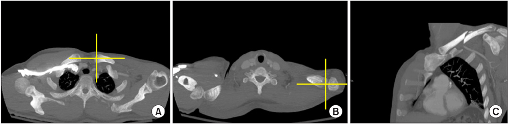
Fig. 2 Depictions of how clavicle length was measured using computed tomography. A clavicle-specific plane was created using Synapse Picture Archiving and Communication System (PACS-Synapse; Fujifilm Medical Systems) software using the center of the proximal end of the clavicle (A) and the distal end of the clavicle (B). (C) The resulting coronal oblique plane image showing the entire length of the clavicle.
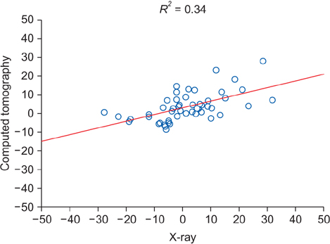
Fig. 3 A linear regression scatter plot of computed tomography length measurements versus X-ray radiographic length measurements. The coefficient of determination (R2) value of 0.34 indicates poor correlation between the two modalities.
Cited by 1 articles
-
A Comparison between Open Reduction/Internal Fixation and Minimally Invasive Plate Osteosynthesis Using a 3-Dimensional Printing Model for Displaced Clavicular Fractures
Dong-Soo Kim, Ho-Seung Jeong, Kyoung-Jin Park, Hyun-Chul Shon, Jae-Young Yang
J Korean Orthop Assoc. 2018;53(4):324-331. doi: 10.4055/jkoa.2018.53.4.324.
Reference
-
1. Marsh JL, Slongo TF, Agel J, et al. Fracture and dislocation classification compendium - 2007: Orthopaedic Trauma Association classification, database and outcomes committee. J Orthop Trauma. 2007; 21:10 Suppl. S1–S133.2. Postacchini F, Gumina S, De Santis P, Albo F. Epidemiology of clavicle fractures. J Shoulder Elbow Surg. 2002; 11(5):452–456.
Article3. Robinson CM. Fractures of the clavicle in the adult: epidemiology and classification. J Bone Joint Surg Br. 1998; 80(3):476–484.4. Neer CS 2nd. Fractures of the distal third of the clavicle. Clin Orthop Relat Res. 1968; 58:43–50.5. Rowe CR. An atlas of anatomy and treatment of midclavicular fractures. Clin Orthop Relat Res. 1968; 58:29–42.6. Eskola A, Vainionpaa S, Myllynen P, Patiala H, Rokkanen P. Outcome of clavicular fracture in 89 patients. Arch Orthop Trauma Surg. 1986; 105(6):337–338.
Article7. Hill JM, McGuire MH, Crosby LA. Closed treatment of displaced middle-third fractures of the clavicle gives poor results. J Bone Joint Surg Br. 1997; 79(4):537–539.
Article8. Lazarides S, Zafiropoulos G. Conservative treatment of fractures at the middle third of the clavicle: the relevance of shortening and clinical outcome. J Shoulder Elbow Surg. 2006; 15(2):191–194.
Article9. Nordqvist A, Petersson CJ, Redlund-Johnell I. Mid-clavicle fractures in adults: end result study after conservative treatment. J Orthop Trauma. 1998; 12(8):572–576.
Article10. Ledger M, Leeks N, Ackland T, Wang A. Short malunions of the clavicle: an anatomic and functional study. J Shoulder Elbow Surg. 2005; 14(4):349–354.
Article11. McKee MD, Pedersen EM, Jones C, et al. Deficits following nonoperative treatment of displaced midshaft clavicular fractures. J Bone Joint Surg Am. 2006; 88(1):35–40.
Article12. Smekal V, Deml C, Irenberger A, et al. Length determination in midshaft clavicle fractures: validation of measurement. J Orthop Trauma. 2008; 22(7):458–462.
Article13. Canadian Orthopaedic Trauma Society. Nonoperative treatment compared with plate fixation of displaced midshaft clavicular fractures: a multicenter, randomized clinical trial. J Bone Joint Surg Am. 2007; 89(1):1–10.14. Robinson CM, Court-Brown CM, McQueen MM, Wakefield AE. Estimating the risk of nonunion following nonoperative treatment of a clavicular fracture. J Bone Joint Surg Am. 2004; 86-A(7):1359–1365.
Article15. McKee RC, Whelan DB, Schemitsch EH, McKee MD. Operative versus nonoperative care of displaced midshaft clavicular fractures: a meta-analysis of randomized clinical trials. J Bone Joint Surg Am. 2012; 94(8):675–684.
Article16. Zlowodzki M, Zelle BA, Cole PA, Jeray K, McKee MD. Evidence-Based Orthopaedic Trauma Working Group. Treatment of acute midshaft clavicle fractures: systematic review of 2144 fractures: on behalf of the Evidence-Based Orthopaedic Trauma Working Group. J Orthop Trauma. 2005; 19(7):504–507.
Article17. Jubel A, Andermahr J, Prokop A, Isenberg J, Rehm KE. Minimal invasive biological osteosynthesis of the clavicle with a titanium nail. Kongressbd Dtsch Ges Chir Kongr. 2002; 119:485–490.18. Jubel A, Andermahr J, Prokop A, Lee JI, Schiffer G, Rehm KE. Treatment of mid-clavicular fractures in adults: early results after rucksack bandage or elastic stable intramedullary nailing. Unfallchirurg. 2005; 108(9):707–714.19. McKee MD, Wild LM, Schemitsch EH. Midshaft malunions of the clavicle: surgical technique. J Bone Joint Surg Am. 2004; 86:Suppl 1. 37–43.20. Walz M, Kolbow B, Auerbach F. Elastic, stable intramedullary nailing in midclavicular fractures: a change in treatment strategies? Unfallchirurg. 2006; 109(3):200–211.
Article21. Jones GL, Bishop JY, Lewis B, Pedroza AD. MOON Shoulder Group. Intraobserver and interobserver agreement in the classification and treatment of midshaft clavicle fractures. Am J Sports Med. 2014; 42(5):1176–1181.
Article22. Bachoura A, Deane AS, Wise JN, Kamineni S. Clavicle morphometry revisited: a 3-dimensional study with relevance to operative fixation. J Shoulder Elbow Surg. 2013; 22(1):e15–e21.
Article23. King PR, Scheepers S, Ikram A. Anatomy of the clavicle and its medullary canal: a computed tomography study. Eur J Orthop Surg Traumatol. 2014; 24(1):37–42.
Article24. Nowak J, Holgersson M, Larsson S. Can we predict longterm sequelae after fractures of the clavicle based on initial findings? A prospective study with nine to ten years of follow-up. J Shoulder Elbow Surg. 2004; 13(5):479–486.
Article25. Matsumura N, Ikegami H, Nakamichi N, et al. Effect of shortening deformity of the clavicle on scapular kinematics: a cadaveric study. Am J Sports Med. 2010; 38(5):1000–1006.
Article26. De Giorgi S, Notarnicola A, Tafuri S, Solarino G, Moretti L, Moretti B. Conservative treatment of fractures of the clavicle. BMC Res Notes. 2011; 4:333.
Article
- Full Text Links
-
- Actions
-
Cited
- CITED
-
- Close
- Share
-
- Similar articles
-
- Arthroscopic Stabilization for Displaced Lateral Clavicular Fractures: Can It Restore Anatomy?
- Computed Tomography in Pelvic Fracture
- Treatment of Clavicle Fracture : Operative vs Non-operative
- Current concepts in the treatment of midshaft clavicle fractures in adults
- Distal Clavicle Fracture in Adolescence Mimicking Type IV Acromioclavicular Joint Injury
