Measurement Methods for Humeral Retroversion Using Two-Dimensional Computed Tomography Scans: Which Is Most Concordant with the Standard Method?
- Affiliations
-
- 1Department of Orthopedic Surgery, Seoul National University College of Medicine, Seoul National University Bundang Hospital, Seongnam, Korea.
- 2Department of Orthopaedic Surgery, Nalgae Hospital, Seoul, Korea. kimw79@naver.com
- 3Department of Orthopaedic Surgery, Cagayan Valley Medical Center, Cagayan, Philippines.
- KMID: 2412293
- DOI: http://doi.org/10.4055/cios.2017.9.2.223
Abstract
- BACKGROUND
Humeral retroversion is variable among individuals, and there are several measurement methods. This study was conducted to compare the concordance and reliability between the standard method and 5 other measurement methods on two-dimensional (2D) computed tomography (CT) scans.
METHODS
CT scans from 21 patients who underwent shoulder arthroplasty (19 women and 2 men; mean age, 70.1 years [range, 42 to 81 years]) were analyzed. The elbow transepicondylar axis was used as a distal reference. Proximal reference points included the central humeral head axis (standard method), the axis of the humeral center to 9 mm posterior to the posterior margin of the bicipital groove (method 1), the central axis of the bicipital groove -30° (method 2), the base axis of the triangular shaped metaphysis +2.5° (method 3), the distal humeral head central axis +2.4° (method 4), and contralateral humeral head retroversion (method 5). Measurements were conducted independently by two orthopedic surgeons.
RESULTS
The mean humeral retroversion was 31.42°± 12.10° using the standard method, and 29.70°± 11.66° (method 1), 30.64°± 11.24° (method 2), 30.41°± 11.17° (method 3), 32.14°± 11.70° (method 4), and 34.15°± 11.47° (method 5) for the other methods. Interobserver reliability and intraobserver reliability exceeded 0.75 for all methods. On the test to evaluate the equality of the standard method to the other methods, the intraclass correlation coefficients (ICCs) of method 2 and method 4 were different from the ICC of the standard method in surgeon A (p < 0.05), and the ICCs of method 2 and method 3 were different form the ICC of the standard method in surgeon B (p < 0.05).
CONCLUSIONS
Humeral version measurement using the posterior margin of the bicipital groove (method 1) would be most concordant with the standard method even though all 5 methods showed excellent agreements.
Keyword
MeSH Terms
Figure
-

Fig. 1 (A) Schematic drawing of the standard method. (B) The transepicondylar axis is defined as a line between the most medial and most lateral extension of the distal humerus. (C) The angle between the central axis of the humeral head and the elbow transepicondylar axis (asterisk) is shown. The central axis is a perpendicular axis of the boundaries of the articular surface determined by using the limits of the subchondral bone in the largest circle of the humeral head.

Fig. 2 (A) Schematic drawing of method 1. (B) The angle between the 9 mm posterior margin of the bicipital groove axis and the elbow transepicondylar axis (asterisk) is shown.
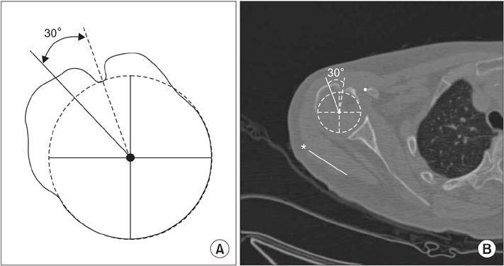
Fig. 3 (A) Schematic drawing of method 2. (B) The angle between the bicipital groove center axis and the elbow transepicondylar axis (asterisk) –30° is shown.
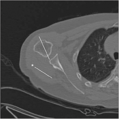
Fig. 4 Method 3 measures the angle between the metaphyseal axis and the elbow transepicondylar axis (asterisk) +2.5°.

Fig. 5 (A) Schematic drawing of method 4. (B) The angle between the axis through the distal humeral head central axis and the elbow transepicondylar axis (asterisk) +2.4° is shown. The distal humeral head central axis is the axis perpendicular to the boundaries of the articular surface determined by using the limits of the subchondral bone on 2 slices (6 mm) below the largest circle of the humeral head on axial slices.
Reference
-
1. Boileau P, Bicknell RT, Mazzoleni N, Walch G, Urien JP. CT scan method accurately assesses humeral head retroversion. Clin Orthop Relat Res. 2008; 466(3):661–669.
Article2. Favre P, Sussmann PS, Gerber C. The effect of component positioning on intrinsic stability of the reverse shoulder arthroplasty. J Shoulder Elbow Surg. 2010; 19(4):550–556.
Article3. Hasan SS, Leith JM, Campbell B, Kapil R, Smith KL, Matsen FA 3rd. Characteristics of unsatisfactory shoulder arthroplasties. J Shoulder Elbow Surg. 2002; 11(5):431–441.
Article4. Pearl ML, Volk AG. Retroversion of the proximal humerus in relationship to prosthetic replacement arthroplasty. J Shoulder Elbow Surg. 1995; 4(4):286–289.
Article5. Krahl VE. An apparatus for measuring the torsion angle in long bones. Science. 1944; 99(2581):498.
Article6. Cyprien JM, Vasey HM, Burdet A, Bonvin JC, Kritsikis N, Vuagnat P. Humeral retrotorsion and glenohumeral relationship in the normal shoulder and in recurrent anterior dislocation (scapulometry). Clin Orthop Relat Res. 1983; (175):8–17.
Article7. Tellioglu AM, Karakas S, Taskin F. Determining torsion angle of humerus head using MRI method. Turk J Med Sci. 2014; 44(4):639–642.
Article8. Ito N, Eto M, Maeda K, Rabbi ME, Iwasaki K. Ultrasonographic measurement of humeral torsion. J Shoulder Elbow Surg. 1995; 4(3):157–161.
Article9. Robertson DD, Yuan J, Bigliani LU, Flatow EL, Yamaguchi K. Three-dimensional analysis of the proximal part of the humerus: relevance to arthroplasty. J Bone Joint Surg Am. 2000; 82(11):1594–1602.
Article10. Hernigou P, Duparc F, Hernigou A. Determining humeral retroversion with computed tomography. J Bone Joint Surg Am. 2002; 84(10):1753–1762.
Article11. Kummer FJ, Perkins R, Zuckerman JD. The use of the bicipital groove for alignment of the humeral stem in shoulder arthroplasty. J Shoulder Elbow Surg. 1998; 7(2):144–146.
Article12. Tillett E, Smith M, Fulcher M, Shanklin J. Anatomic determination of humeral head retroversion: the relationship of the central axis of the humeral head to the bicipital groove. J Shoulder Elbow Surg. 1993; 2(5):255–256.
Article13. Athwal GS, MacDermid JC, Goel DP. Metaversion can reliably predict humeral head version: a computed tomography-based validation study. J Shoulder Elbow Surg. 2010; 19(8):1145–1149.
Article14. DeLude JA, Bicknell RT, MacKenzie GA, et al. An anthropometric study of the bilateral anatomy of the humerus. J Shoulder Elbow Surg. 2007; 16(4):477–483.
Article15. Farrokh D, Fabeck L, Descamps PY, Hardy D, Delince P. Computed tomography measurement of humeral head retroversion: influence of patient positioning. J Shoulder Elbow Surg. 2001; 10(6):550–553.
Article16. Shrout PE, Fleiss JL. Intraclass correlations: uses in assessing rater reliability. Psychol Bull. 1979; 86(2):420–428.
Article17. Donner A, Shoukri MM, Klar N, Bartfay E. Testing the equality of two dependent kappa statistics. Stat Med. 2000; 19(3):373–387.
Article18. Dashottar A, Borstad JD. Validity of measuring humeral torsion using palpation of bicipital tuberosities. Physiother Theory Pract. 2013; 29(1):67–74.
Article19. Harrold F, Wigderowitz C. A three-dimensional analysis of humeral head retroversion. J Shoulder Elbow Surg. 2012; 21(5):612–617.
Article
- Full Text Links
-
- Actions
-
Cited
- CITED
-
- Close
- Share
-
- Similar articles
-
- Little Leaguer's Shoulder Can Cause Severe Three-Dimensional Humeral Deformity
- Measurement of Proximal Humerus in Korean Adult Skeleton
- Normal Humeral Head Retroversion Angle in Korean Measured with Semil - axial View
- Evaluation of the accuracy and reliability of 3-dimensional computerized tomography for measurement of maxillofacial region
- Comparative Analysis of Accuracy between Computerized Tomography and Cephalogram for 3-Dimensional Measurement of Maxillofacial Structure
