Comparison of the Extent of Degeneration among the Normal Disc, Immobilized Disc, and Immobilized Disc with an Endplate Fracture
- Affiliations
-
- 1Department of Orthopaedic Surgery, Daegu Catholic University Medical Center, Daegu, Korea. bong@cu.ac.kr
- KMID: 2412288
- DOI: http://doi.org/10.4055/cios.2017.9.2.193
Abstract
- BACKGROUND
This study attempts to prove a cause and effect relationship between spine immobilization following posterior fixation for unstable burst fractures and degeneration observed following hardware removal.
METHODS
We enrolled 57 patients (259 intervertebral discs [IVDs]) who underwent only posterior instrumentation without fusion for thoracolumbar and lumbar unstable burst fractures. We arbitrarily named the IVD that has an endplate fracture after immobilization using pedicle screws as the fractured endplate and immobilized disc (FEID), the IVD that has no endplate fracture after immobilization using pedicle screws as the nonfractured endplate and immobilized disc (NFEID), and the IVD that has no endplate fracture and no immobilization instrumentation as the normal disc (ND). At 2 years after implant removal, magnetic resonance imaging (MRI) was performed again for comparison. The extent of disc degeneration was classified using the Pfirrmann classification system.
RESULTS
FEIDs were present in 67 levels, NFEIDs in 78 levels, and NDs in 114 levels. According to the Pfirrmann classification, 7.9% of the NDs, 32.1% of the NFEIDs, and 43.3% of the FEIDs were more degenerated at 2 years after implant removal. The FEIDs and NFEIDs were more degenerated than the NDs and the FEIDs were more degenerated than the NFEIDs at statistically significant levels (p < 0.001 for both).
CONCLUSIONS
Spine immobilization with transpedicular screws has a significant influence on disc degeneration, and an endplate fracture accelerates the degeneration process.
MeSH Terms
-
Adolescent
Adult
Cohort Studies
Female
Fracture Fixation, Internal/*statistics & numerical data
Humans
*Intervertebral Disc/diagnostic imaging/pathology/surgery
*Intervertebral Disc Degeneration/diagnostic imaging/pathology
Magnetic Resonance Imaging
Male
Middle Aged
Pedicle Screws
*Spinal Fractures/diagnostic imaging/pathology/surgery
Young Adult
Figure
-
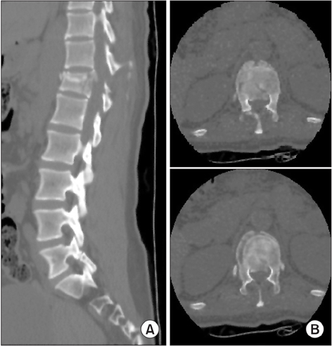
Fig. 1 Displaced endplate fracture was diagnosed based on sagittal (A) and coronal (B) images of computed tomography.
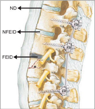
Fig. 2 Intervertebral discs (IVDs) after instrumentation for spine fracture. ND: normal disc, an IVD that has no endplate fracture and no immobilization. NFEID: nonfractured endplate and immobilized disc, an IVD that has no endplate fracture but is immobilized using pedicle screws. FEID: fractured endplate and immobilized disc, an IVD that has endplate fracture and is immobilized using pedicle screws. *Fractured vertebra.
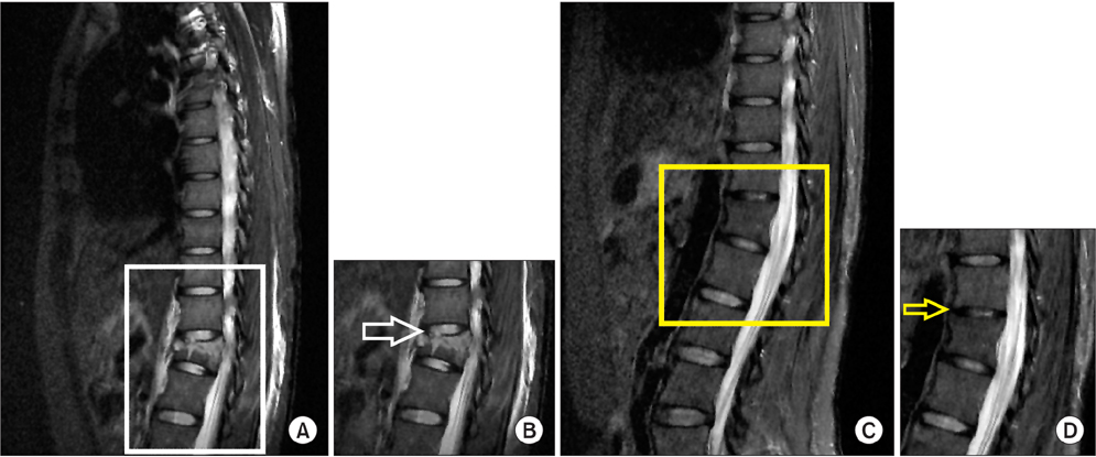
Fig. 3 Serial magnetic resonance imaging (MRI) showing more degenerative changes. (A) MRI imaging of the fractured vertebral body and injured endplate (white square) at the time of injury shows grade 1 degeneration. (B) Disc is homogeneous with bright hyperintense white signal intensity and normal disc height (white arrow). (C) MRI imaging of the treated vertebral body and endplate (yellow square) at 2 years after removal surgery shows grade 3 degeneration. (D) Disc is inhomogenous with an intermittent gray signal intensity and disc height is slightly or moderately decreased (yellow arrow).
Reference
-
1. Moore RJ. The vertebral endplate: disc degeneration, disc regeneration. Eur Spine J. 2006; 15:Suppl 3. S333–S337.
Article2. Edwards WT, Zheng Y, Ferrara LA, Yuan HA. Structural features and thickness of the vertebral cortex in the thoracolumbar spine. Spine (Phila Pa 1976). 2001; 26(2):218–225.
Article3. Roberts S, Menage J, Urban JP. Biochemical and structural properties of the cartilage end-plate and its relation to the intervertebral disc. Spine (Phila Pa 1976). 1989; 14(2):166–174.
Article4. Vernon-Roberts B. Age-related and degenerative pathology of intervertebral discs and apophyseal joints. In : Jayson MI, editor. The lumbar spine and back pain. 4th ed. Edinburgh, Scotland: Churchill Livingstone;1992. p. 17–41.5. Ghanem N, Uhl M, Muller C, et al. MRI and discography in traumatic intervertebral disc lesions. Eur Radiol. 2006; 16(11):2533–2541.
Article6. Oner FC, van Gils AP, Dhert WJ, Verbout AJ. MRI findings of thoracolumbar spine fractures: a categorisation based on MRI examinations of 100 fractures. Skeletal Radiol. 1999; 28(8):433–443.
Article7. Kirkaldy-Willis WH, Wedge JH, Yong-Hing K, Reilly J. Pathology and pathogenesis of lumbar spondylosis and stenosis. Spine (Phila Pa 1976). 1978; 3(4):319–328.
Article8. Stokes IA, Iatridis JC. Mechanical conditions that accelerate intervertebral disc degeneration: overload versus immobilization. Spine (Phila Pa 1976). 2004; 29(23):2724–2732.
Article9. Pfirrmann CW, Metzdorf A, Zanetti M, Hodler J, Boos N. Magnetic resonance classification of lumbar intervertebral disc degeneration. Spine (Phila Pa 1976). 2001; 26(17):1873–1878.
Article10. Taylor JR. Growth of human intervertebral discs and vertebral bodies. J Anat. 1975; 120(Pt 1):49–68.11. Holm S, Maroudas A, Urban JP, Selstam G, Nachemson A. Nutrition of the intervertebral disc: solute transport and metabolism. Connect Tissue Res. 1981; 8(2):101–119.
Article12. Knop C, Blauth M, Buhren V, et al. Surgical treatment of injuries of the thoracolumbar transition. 3: follow-up examination. Results of a prospective multi-center study by the “Spinal” Study Group of the German Society of Trauma Surgery. Unfallchirurg. 2001; 104(7):583–600.13. Haschtmann D, Stoyanov JV, Gedet P, Ferguson SJ. Vertebral endplate trauma induces disc cell apoptosis and promotes organ degeneration in vitro. Eur Spine J. 2008; 17(2):289–299.
Article14. Malcolm BW, Bradford DS, Winter RB, Chou SN. Post-traumatic kyphosis: a review of forty-eight surgically treated patients. J Bone Joint Surg Am. 1981; 63(6):891–899.
Article15. Denis F. The three column spine and its significance in the classification of acute thoracolumbar spinal injuries. Spine (Phila Pa 1976). 1983; 8(8):817–831.
Article16. Lindsey RW, Dick W. The fixateur interne in the reduction and stabilization of thoracolumbar spine fractures in patients with neurologic deficit. Spine (Phila Pa 1976). 1991; 16:3 Suppl. S140–S145.
Article17. Steindl A, Schuh G. Late results after lumbar vertebrae fracture with Lorenz Bohler conservative treatment. Unfallchirurg. 1992; 95(9):439–444.18. Akbarnia BA, Crandall DG, Burkus K, Matthews T. Use of long rods and a short arthrodesis for burst fractures of the thoracolumbar spine: a long-term follow-up study. J Bone Joint Surg Am. 1994; 76(11):1629–1635.
Article19. Speth MJ, Oner FC, Kadic MA, de Klerk LW, Verbout AJ. Recurrent kyphosis after posterior stabilization of thoracolumbar fractures: 24 cases treated with a Dick internal fixator followed for 1.5-4 years. Acta Orthop Scand. 1995; 66(5):406–410.
Article20. Oner FC, van der Rijt RR, Ramos LM, Dhert WJ, Verbout AJ. Changes in the disc space after fractures of the thoracolumbar spine. J Bone Joint Surg Br. 1998; 80(5):833–839.
Article21. Boos N, Weissbach S, Rohrbach H, Weiler C, Spratt KF, Nerlich AG. Classification of age-related changes in lumbar intervertebral discs: 2002 Volvo Award in basic science. Spine (Phila Pa 1976). 2002; 27(23):2631–2644.22. Cheung KM, Karppinen J, Chan D, et al. Prevalence and pattern of lumbar magnetic resonance imaging changes in a population study of one thousand forty-three individuals. Spine (Phila Pa 1976). 2009; 34(9):934–940.
Article23. Lin RM, Panjabi MM, Oxland TR. Functional radiographs of acute thoracolumbar burst fractures: a biomechanical study. Spine (Phila Pa 1976). 1993; 18(16):2431–2437.24. Ching CT, Chow DH, Yao FY, Holmes AD. The effect of cyclic compression on the mechanical properties of the inter-vertebral disc: an in vivo study in a rat tail model. Clin Biomech (Bristol, Avon). 2003; 18(3):182–189.
Article25. Kumar MN, Jacquot F, Hall H. Long-term follow-up of functional outcomes and radiographic changes at adjacent levels following lumbar spine fusion for degenerative disc disease. Eur Spine J. 2001; 10(4):309–313.
Article26. Roaf R. A study of the mechanics of spinal injuries. J Bone Joint Surg Br. 1960; 42(4):810–823.
Article27. Modic MT, Ross JS. Lumbar degenerative disk disease. Radiology. 2007; 245(1):43–61.
Article28. Boden SD, Davis DO, Dina TS, Patronas NJ, Wiesel SW. Abnormal magnetic-resonance scans of the lumbar spine in asymptomatic subjects: a prospective investigation. J Bone Joint Surg Am. 1990; 72(3):403–408.
Article29. Sato H, Kikuchi S. The natural history of radiographic instability of the lumbar spine. Spine (Phila Pa 1976). 1993; 18(14):2075–2079.
Article
- Full Text Links
-
- Actions
-
Cited
- CITED
-
- Close
- Share
-
- Similar articles
-
- The Effect of Disc Degeneration in Osteoporotic Vertebral Fracture
- Biochemical Factors of Intervertebral Disc Degeneration: Implications for Disc Regeneration
- Histological Composition of the Extruded Intervertebral Disc in Lumbar Intervertebral Disc Herniation
- Changes of Posterior Bulging of the Lumbar Intervertebral Discs with Flexion and Extension in Central Disc Bulges and Disc Degeneration
- Lumbar Disc Degeneration and Segmental Instability: A Comparison of Magnetic Resonance Images and Plain Radiographs
