Rotational Acetabular Osteotomy
- Affiliations
-
- 1Department of Orthopaedic Surgery, Hiroshima Prefectural Rehabilitation Center, Higashi-Hiroshima, Japan. yasuyuji@hiroshima-wsc.jp
- 2Department of Orthopaedic Surgery, Mihara Medical Association Hospital, Hiroshima, Japan.
- 3Department of Artificial Joints and Biomaterials, Hiroshima University, Higashi-Hiroshima, Japan.
- 4Department of Orthopaedic Surgery, Hiroshima University, Higashi-Hiroshima, Japan.
- 5Hiroshima University, Higashi-Hiroshima, Japan.
- KMID: 2412279
- DOI: http://doi.org/10.4055/cios.2017.9.2.129
Abstract
- Hip dysplasia is the most common cause of secondary osteoarthritis (OA). To prevent the early onset of secondary OA, Nishio's transposition osteotomy, Steel's triple osteotomy, Eppright's dial osteotomy, Wagner's spherical acetabular osteotomy, Tagawa's rotational acetabular osteotomy (RAO), and Ganz' periacetabular osteotomy (PAO) have been proposed. PAO and RAO are now commonly used in surgical treatment of symptomatic acetabular dysplasia in Europe, North America, and Asia. The aim of this paper is to present the followings: the patient selection criteria for RAO; the surgical technique of RAO; the long-term outcome of RAO; and the future perspectives.
Keyword
MeSH Terms
Figure
-
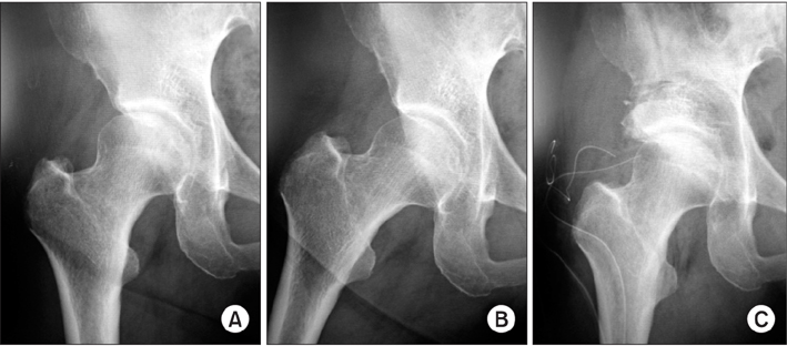
Fig. 1 Preoperative and postoperative radiographs. Preoperative anteroposterior (AP) views in neutral position (A) and abducted position (B). (C) Postoperative AP view in neutral position.
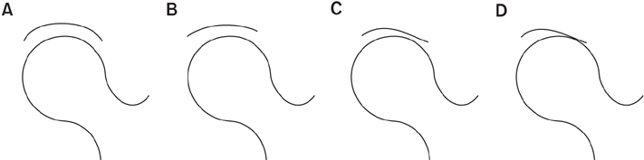
Fig. 2 Postoperative joint congruency is classified into 4 grades: excellent (A), good (B), fair (C), and poor (D).
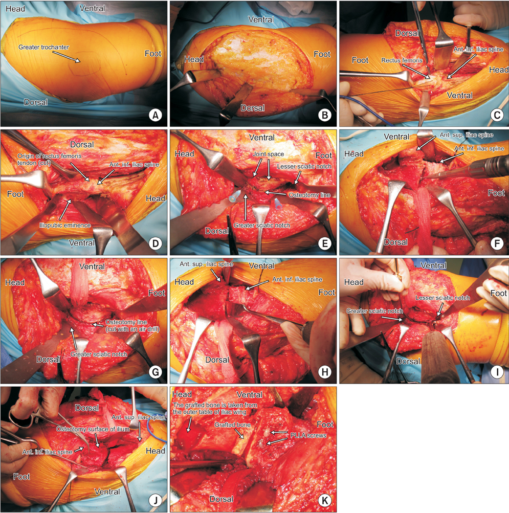
Fig. 3 Surgical technique. (A) Skin incision. (B) The skin flap is elevated in posterior direction. (C) The rectus femoris is cut. (D) The site for osteotomy of the pubic bone is exposed. (E) The osteotomy line of the posterior wall of the acetabulum. A needle has been inserted in the joint. (F) The outer cortical bone of the ilium is cut with an air drill. (G) The outer cortical bone of the posterior wall and ischium has been cut. (H) The osteotomy of the anterior part of the ilium is shown. (I) The osteotomy of the posterior wall and ischium is shown. (J) The acetabulum is rotated in anterolateral direction. (K) The acetabulum is fixed with two poly-L-lactic acid (PLLA) screws. Ant: anterior, Inf: inferior, Sup: superior.
Cited by 2 articles
-
Pelvic Osteotomy in Adults
Yoon-Je Cho, Young-Wan Ko
J Korean Orthop Assoc. 2017;52(6):500-513. doi: 10.4055/jkoa.2017.52.6.500.Radiographic Progression of Osteoarthritis after Rotational Acetabular Osteotomy: Minimum 10-Year Follow-up Outcome According to the Tönnis Grade
Byung-Woo Min, Chang Soo Kang, Kyung-Jae Lee, Ki-Cheor Bae, Chul-Hyun Cho, Jung-Hoon Choi, Hyuk-Joon Sohn, Hong-Kwan Sin
Clin Orthop Surg. 2018;10(3):299-306. doi: 10.4055/cios.2018.10.3.299.
Reference
-
1. Aronson J. Osteoarthritis of the young adult hip: etiology and treatment. Instr Course Lect. 1986; 35:119–128.2. Harris WH. Etiology of osteoarthritis of the hip. Clin Orthop Relat Res. 1986; (213):20–33.
Article3. Murphy SB, Ganz R, Muller ME. The prognosis in untreated dysplasia of the hip: a study of radiographic factors that predict the outcome. J Bone Joint Surg Am. 1995; 77(7):985–989.
Article4. Nishio A. Transposition osteotomy of the acetabulum for the treatment of congenital dislocation of the hip. Nippon Seikeigeka Gakkai Zasshi. 1956; 30:482–484.5. Steel HH. Triple osteotomy of the innominate bone. J Bone Joint Surg Am. 1973; 55(2):343–350.
Article6. Eppright RH. Dial osteotomy of the acetabulum in the treatment of dysplasia of the hip. J Bone Joint Surg Am. 1975; 57(8):1172.7. Wagner H. Osteotomies for congenital hip dislocation. The Hip Society. The hip: proceedings of the fourth open scientific meeting of the Hip Society. St Louis, MO: C.V. Mosby;1976. p. 45–66.8. Ninomiya S, Tagawa H. Rotational acetabular osteotomy for the dysplastic hip. J Bone Joint Surg Am. 1984; 66(3):430–436.
Article9. Ganz R, Klaue K, Vinh TS, Mast JW. A new periacetabular osteotomy for the treatment of hip dysplasias: technique and preliminary results. Clin Orthop Relat Res. 1988; (232):26–36.
Article10. Leunig M, Siebenrock KA, Ganz R. Rationale of periacetabular osteotomy and background work. Instr Course Lect. 2001; 50:229–238.
Article11. Clohisy JC, Schutz AL, St John L, Schoenecker PL, Wright RW. Periacetabular osteotomy: a systematic literature review. Clin Orthop Relat Res. 2009; 467(8):2041–2052.
Article12. Yasunaga Y, Yamasaki T, Ochi M. Patient selection criteria for periacetabular osteotomy or rotational acetabular osteotomy. Clin Orthop Relat Res. 2012; 470(12):3342–3354.
Article13. Trousdale RT, Ekkernkamp A, Ganz R, Wallrichs SL. Periacetabular and intertrochanteric osteotomy for the treatment of osteoarthrosis in dysplastic hips. J Bone Joint Surg Am. 1995; 77(1):73–85.
Article14. Nakamura S, Ninomiya S, Takatori Y, Morimoto S, Umeyama T. Long-term outcome of rotational acetabular osteotomy: 145 hips followed for 10-23 years. Acta Orthop Scand. 1998; 69(3):259–265.
Article15. Siebenrock KA, Scholl E, Lottenbach M, Ganz R. Bernese periacetabular osteotomy. Clin Orthop Relat Res. 1999; (363):9–20.
Article16. Yasunaga Y, Ochi M, Terayama H, Tanaka R, Yamasaki T, Ishii Y. Rotational acetabular osteotomy for advanced osteoarthritis secondary to dysplasia of the hip. J Bone Joint Surg Am. 2006; 88(9):1915–1919.
Article17. Hasegawa Y, Kanoh T, Seki T, Matsuoka A, Kawabe K. Joint space wider than 2 mm is essential for an eccentric rotational acetabular osteotomy for adult hip dysplasia. J Orthop Sci. 2010; 15(5):620–625.
Article18. Millis MB, Kain M, Sierra R, et al. Periacetabular osteotomy for acetabular dysplasia in patients older than 40 years: a preliminary study. Clin Orthop Relat Res. 2009; 467(9):2228–2234.
Article19. Yasunaga Y, Takahashi K, Ochi M, et al. Rotational acetabular osteotomy in patients forty-six years of age or older: comparison with younger patients. J Bone Joint Surg Am. 2003; 85(2):266–272.
Article20. Yamaguchi J, Hasegawa Y, Kanoh T, Seki T, Kawabe K. Similar survival of eccentric rotational acetabular osteotomy in patients younger and older than 50 years. Clin Orthop Relat Res. 2009; 467(10):2630–2637.
Article21. Kaneuji A, Sugimori T, Ichiseki T, Fukui K, Takahashi E, Matsumoto T. Rotational acetabular osteotomy for osteoarthritis with acetabular dysplasia: conversion rate to total hip arthroplasty within twenty years and osteoarthritis progression after a minimum of twenty years. J Bone Joint Surg Am. 2015; 97(9):726–732.
Article22. Hasegawa Y, Iwase T, Kitamura S, Kawasaki M, Yamaguchi J. Eccentric rotational acetabular osteotomy for acetabular dysplasia and osteoarthritis: follow-up at a mean duration of twenty years. J Bone Joint Surg Am. 2014; 96(23):1975–1982.
Article23. Yasunaga Y, Ochi M, Yamasaki T, Shoji T, Izumi S. Rotational acetabular osteotomy for pre- and early osteoarthritis secondary to dysplasia provides durable results at 20 years. Clin Orthop Relat Res. 2016; 474(10):2145–2153.
Article24. Kain MS, Novais EN, Vallim C, Millis MB, Kim YJ. Periacetabular osteotomy after failed hip arthroscopy for labral tears in patients with acetabular dysplasia. J Bone Joint Surg Am. 2011; 93:Suppl 2. 57–61.
Article25. Kim SD, Jessel R, Zurakowski D, Millis MB, Kim YJ. Anterior delayed gadolinium-enhanced MRI of cartilage values predict joint failure after periacetabular osteotomy. Clin Orthop Relat Res. 2012; 470(12):3332–3341.
Article26. Millis MB. Congenital hip dysplasia: treatment from infancy to skeletal maturity. In : Tronzo RG, editor. Surgery of the hip joint. 2nd ed. Vol. 1. New York, NY: Springer;1984. p. 329–385.27. Millis MB, Murphy SB, Poss R. Osteotomies about the hip for the prevention and treatment of osteoarthrosis. Instr Course Lect. 1996; 45:209–226.
Article28. Shimogaki K, Yasunaga Y, Ochi M. A histological study of articular cartilage after rotational acetabular osteotomy for hip dysplasia. J Bone Joint Surg Br. 2005; 87(7):1019–1023.
Article29. Yasunaga Y, Ikuta Y, Kanazawa T, Takahashi K, Hisatome T. The state of the articular cartilage at the time of surgery as an indication for rotational acetabular osteotomy. J Bone Joint Surg Br. 2001; 83(7):1001–1004.
Article30. Kim YJ, Jaramillo D, Millis MB, Gray ML, Burstein D. Assessment of early osteoarthritis in hip dysplasia with delayed gadolinium-enhanced magnetic resonance imaging of cartilage. J Bone Joint Surg Am. 2003; 85(10):1987–1992.
Article31. Locher S, Werlen S, Leunig M, Ganz R. MR-arthrography with radial sequences for visualization of early hip pathology not visible on plain radiographs. Z Orthop Ihre Grenzgeb. 2002; 140(1):52–57.32. Watanabe A, Boesch C, Siebenrock K, Obata T, Anderson SE. T2 mapping of hip articular cartilage in healthy volunteers at 3T: a study of topographic variation. J Magn Reson Imaging. 2007; 26(1):165–171.
Article33. Cunningham T, Jessel R, Zurakowski D, Millis MB, Kim YJ. Delayed gadolinium-enhanced magnetic resonance imaging of cartilage to predict early failure of Bernese periacetabular osteotomy for hip dysplasia. J Bone Joint Surg Am. 2006; 88(7):1540–1548.
Article34. Garbuz DS, Awwad MA, Duncan CP. Periacetabular osteotomy and total hip arthroplasty in patients older than 40 years. J Arthroplasty. 2008; 23(7):960–963.
Article35. Hsieh PH, Huang KC, Lee PC, Chang YH. Comparison of periacetabular osteotomy and total hip replacement in the same patient: a two- to ten-year follow-up study. J Bone Joint Surg Br. 2009; 91(7):883–888.36. Sharifi E, Sharifi H, Morshed S, Bozic K, Diab M. Cost-effectiveness analysis of periacetabular osteotomy. J Bone Joint Surg Am. 2008; 90(7):1447–1456.
Article37. Teratani T, Naito M, Kiyama T, Maeyama A. Periacetabular osteotomy in patients fifty years of age or older. J Bone Joint Surg Am. 2010; 92(1):31–41.
Article38. van Bergayk AB, Garbuz DS. Quality of life and sports-specific outcomes after Bernese periacetabular osteotomy. J Bone Joint Surg Br. 2002; 84(3):339–343.
Article39. McAuley JP, Szuszczewicz ES, Young A, Engh CA Sr. Total hip arthroplasty in patients 50 years and younger. Clin Orthop Relat Res. 2004; (418):119–125.
Article40. Haviv B, Singh PJ, Takla A, O'Donnell J. Arthroscopic femoral osteochondroplasty for cam lesions with isolated acetabular chondral damage. J Bone Joint Surg Br. 2010; 92(5):629–633.
Article41. Yen YM, Kocher MS. Chondral lesions of the hip: microfracture and chondroplasty. Sports Med Arthrosc. 2010; 18(2):83–89.42. Pridie KH. A method of resurfacing osteoarthritic knee joints. J Bone Joint Surg Br. 1959; 41(3):618–619.
- Full Text Links
-
- Actions
-
Cited
- CITED
-
- Close
- Share
-
- Similar articles
-
- Pelvic Osteotomy in Adults
- Rotational Acetabular Osteotomy for the Dysplastic Acetabulum
- Rotational Acetabular Osteotomy for the Dysplastic Hip: A Follow-up for 5 to 18 years
- Rotational acetabular osteotomy in acetabular dysplasia
- Stress Distribution According to Center-Edge Angle in Dysplastic Hip and the Biomechanical Effect of Rotational Acetabular Osteotomy
