A Rare Case of Intra-Articular Displacement of EndoButton Following Anterior Cruciate Ligament Reconstruction
- Affiliations
-
- 1Department of Orthopaedic Surgery, Tan Tock Seng Hospital, Singapore. sean.ho@mohh.com.sg
- KMID: 2412256
- DOI: http://doi.org/10.4055/cios.2017.9.4.534
Abstract
- The EndoButton is a commonly used device for femoral fixation of anterior cruciate ligament grafts. Complications from its usage remain rare. Incorrect femoral tunnel placement may increase the risk of intra-articular displacement of the EndoButton. We present a case of anterior femoral tunnel placement resulting in intra-articular displacement of the EndoButton after failure. A 24-year-old man presented to us after failure of anterior cruciate ligament reconstruction performed 3 years prior. Radiographs revealed an intra-articular displacement of the EndoButton. Intraoperatively, it was noted that the femoral tunnel exit was within the suprapatellar pouch, with the displaced EndoButton lodged between the posterior aspect of the lateral tibial plateau and the capsule. Intra-articular displacement of the EndoButton is a rare complication and has only been reported twice in the literature. Anterior placement of the femoral tunnel may predispose patients to this complication and it is recommended to check the EndoButton position intraoperatively to avoid such a complication, especially for the unexperienced surgeon.
MeSH Terms
Figure
-
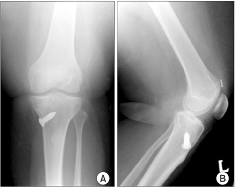
Fig. 1 Anteroposterior (A) and lateral (B) X-rays of the left knee after initial anterior cruciate ligament reconstruction. The EndoButton (Smith & Nephew) is in an anterior position within the suprapatellar pouch.
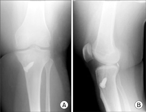
Fig. 2 Anteroposterior (A) and lateral (B) X-rays of the left knee showing a displaced intra-articular EndoButton (Smith & Nephew) within the popliteal space.

Fig. 3 T1-weighted magnetic resonance imaging of the left knee showing an indistinct anterior cruciate ligament graft (white arrow).
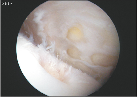
Fig. 4 Intraoperative arthroscopic view showing a frayed continuous loop of the EndoButton (Smith & Nephew).
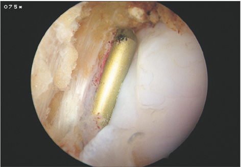
Fig. 5 Intraoperative arthroscopic view of the retained EndoButton (Smith & Nephew). The button was lodged posterior to the lateral tibial plateau.
Reference
-
1. Ahmad CS, Gardner TR, Groh M, Arnouk J, Levine WN. Mechanical properties of soft tissue femoral fixation devices for anterior cruciate ligament reconstruction. Am J Sports Med. 2004; 32(3):635–640.
Article2. Muneta T, Yagishita K, Kurihara Y, Sekiya I. Intra-articular detachment of the Endobutton more than 18 months after anterior cruciate ligament reconstruction. Arthroscopy. 1999; 15(7):775–778.
Article3. Yanmis I, Tunay S, Oguz E, Yildiz C, Ozkan H, Kirdemir V. Dropping of an EndoButton into the knee joint 2 years after anterior cruciate ligament repair using proximal fixation methods. Arthroscopy. 2004; 20(6):641–643.
Article4. Bylski-Austrow DI, Grood ES, Hefzy MS, Holden JP, Butler DL. Anterior cruciate ligament replacements: a mechanical study of femoral attachment location, flexion angle at tensioning, and initial tension. J Orthop Res. 1990; 8(4):522–531.
Article5. Zysk SP, Fraunberger P, Veihelmann A, et al. Tunnel enlargement and changes in synovial fluid cytokine profile following anterior cruciate ligament reconstruction with patellar tendon and hamstring tendon autografts. Knee Surg Sports Traumatol Arthrosc. 2004; 12(2):98–103.
Article
- Full Text Links
-
- Actions
-
Cited
- CITED
-
- Close
- Share
-
- Similar articles
-
- Ligament Reconstruction in Congenital Absence of the Anterior Cruciate Ligament: A Case Report
- Arthroscopic ACL Reconstruction with Quadrupled Semitendinosus Tendon and endobutton
- Anterior Cruciate Ligament Reconstruction with Achilles Tendon Allograft
- The Effect of Cyclic Tensile Load on Various Tibial Fixation Techniques in Anterior Cruciate Ligament Reconstruction
- Open anterior cruciate ligament reconstruction using inside-out femoral drilling
