Congenital Absence of the Left Atrial Appendage: An Unexpected and Incidental Anomaly in a Patient with Multiple Cerebellar Infarctions
- Affiliations
-
- 1Division of Cardiology, Department of Internal Medicine, College of Medicine, Dankook University, Cheonan, Korea. kdongmin@dankook.ac.kr
- KMID: 2411898
- DOI: http://doi.org/10.4068/cmj.2018.54.2.133
Abstract
- No abstract available.
MeSH Terms
Figure
-
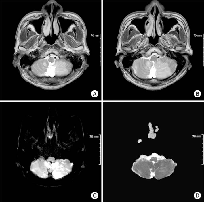
FIG. 1 T2-weighted (A, B), diffusion (C) and apparent diffusion coefficient (D) brain magnetic resonance imaging of the patient reveals acute infarctions in both cerebellums associated with the posteroinferior cellebellar artery (left dominant).
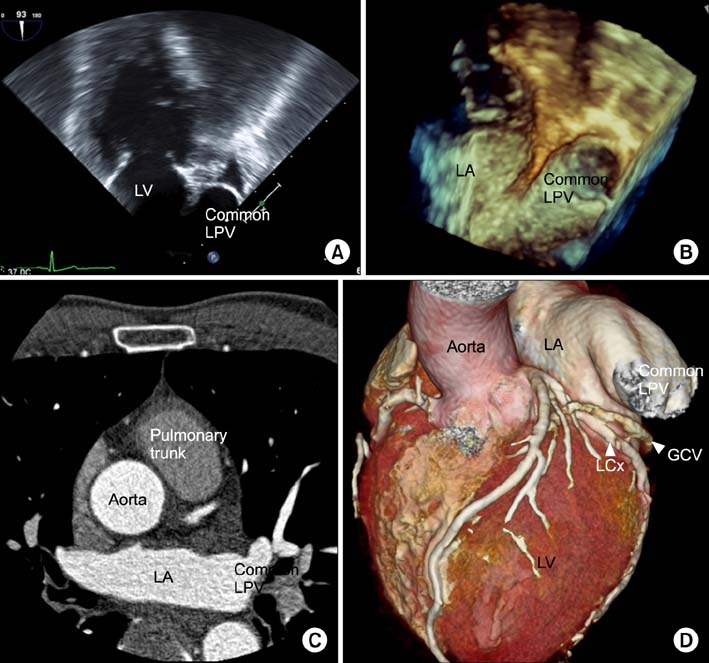
FIG. 2 Multiple transesophageal echocardiography image views at the midesophageal level (A, B). It was impossible to visualize the left atrial appendage in its typical location. Contrast-enhanced multidetector computed tomography with axial (C) and 3 dimensional volume-rendering (D) images. The left atrial appendage was not visualized. In contrast, the great cardiac vein and left circumflex coronary artery were clearly observed along with the atrioventricular groove. LA: left atrium, LV: left ventricle, LPV: left pulmonary vein, GCV: great cardiac vein, LCx: left circumflex coronary artery.
Reference
-
1. Collier P, Cavalcante JL, Phelan D, Thavendiranathan P, Dahiya A, Grant A, et al. Congenital absence of the left atrial appendage. Circ Cardiovasc Imaging. 2012; 5:549–550.
Article2. Zhang ZJ, Dong JZ, Ma CS. Congenital absence of the left atrial appendage: a rare anatomical variation with clinical significance. Acta Cardiol. 2013; 68:325–327.
Article3. Song IG, Kim SH, Oh YS, Rho TH. Underdevelopment of left atrial appendage. Korean Circ J. 2017; 47:141–143.
Article
- Full Text Links
-
- Actions
-
Cited
- CITED
-
- Close
- Share
-
- Similar articles
-
- Resection of a Congenital Left Atrial Appendage Aneurysm without Extracorporeal Circulation
- Congenital Left Atrial Appendage Aneurysm: A Case Report
- Underdevelopment of Left Atrial Appendage
- Confirmation of absence: the need for advanced imaging of the left atrial appendage
- Persistent Atrial Fibrillation Related to a Congenital Pericardial Defect and Left Atrial Appendage Herniation
