Intraosseous Lipoma: 18 Years of Experience at a Single Institution
- Affiliations
-
- 1Department of Orthopedic Surgery, Kosin University Gospel Hospital, Busan, Korea. shchung@kosin.ac.kr
- 2Department of Pathology, Kosin University Gospel Hospital, Busan, Korea.
- 3Department of Radiology, Kosin University Gospel Hospital, Busan, Korea.
- KMID: 2411750
- DOI: http://doi.org/10.4055/cios.2018.10.2.234
Abstract
- BACKGROUND
Intraosseous lipoma is a very rare lesion that constitutes no more than 0.1% of all bone tumors. We analyzed 21 cases of intraosseous lipoma at a single institution for clinical and radiographic characteristics.
METHODS
A retrospective study was performed on 21 pathologically confirmed intraosseous lipomas treated in our hospital from 2000 to 2017. Simple X-ray and magnetic resonance imaging findings and medical records were reviewed. Patients' age, sex, and clinical symptoms were investigated. From the radiographic images, the site of the lesion, calcification, bony expansion, and stage of the lesion were evaluated. Correlations between the degree of involution and clinical symptoms were analyzed.
RESULTS
The mean age of patients was 50 years (range, 20 to 67 years), and there were 13 males and eight females. The mean lesion size was 6.1 cm (range, 2.5 to 13.6 cm). The most common anatomical site of the lesion was the femur (seven cases), and three cases occurred in flat bones such as the ilium and scapula. Visual analogue scale score for pain was 3 to 6 in 15 patients. There were no complaints of functional limitation. There was no correlation between the degree of degeneration and clinical symptoms (p = 1.000). Curettage was performed as a surgical treatment in 20 patients, and bone graft was performed using a bone chip. Excision was performed in one patient. Pain was resolved in seven of 11 patients with a complaint of preoperative pain; intermittent pain remained in four cases. There was no local recurrence or malignant change during the follow-up.
CONCLUSIONS
There was no correlation between the degree of degeneration and clinical symptoms. Pain was the most common clinical symptom, but it was rarely accompanied by functional limitation. However, it is important to distinguish it from other pain-inducing disorders. The incidence of intraosseous lipomas is low, and detection based on various imaging findings can be difficult. Clear understanding of the radiographic findings and symptoms of intraosseous lipoma is helpful for diagnosis and differentiation.
Keyword
MeSH Terms
Figure
-
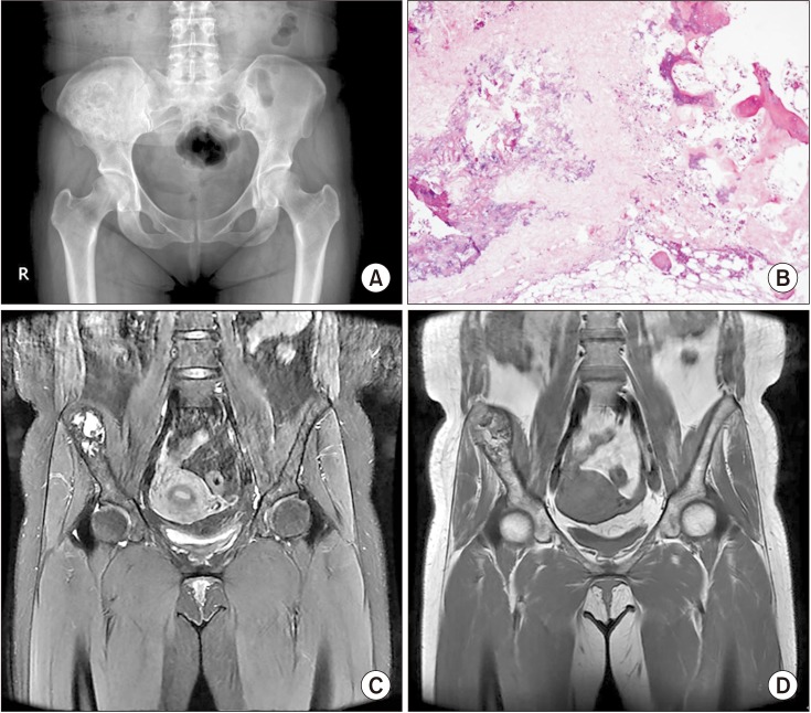
Fig. 1 A 44-year-old woman with an intraosseous lipoma (late phase) in the right ilium. (A) The anteroposterior pelvic radiograph shows a sclerotic-margined and expansile bone lesion in the right iliac bone. (B) Low power photomicrograph demonstrating fat necrosis with fine, powdery calcification and fibrosis, similar to what is seen in bone infarct. These features represent the late stage intraosseous lipoma (H&E, ×100). (C) On fat-suppressed coronal T2-weighted magnetic resonance imaging, the mass has high signal intensity centrally, indicating cystic necrotic portions. (D) On coronal T1-weighted magnetic resonance imaging, the mass demonstrates well-defined fat signal intensity with a central area of low signal intensity due to cystic change.
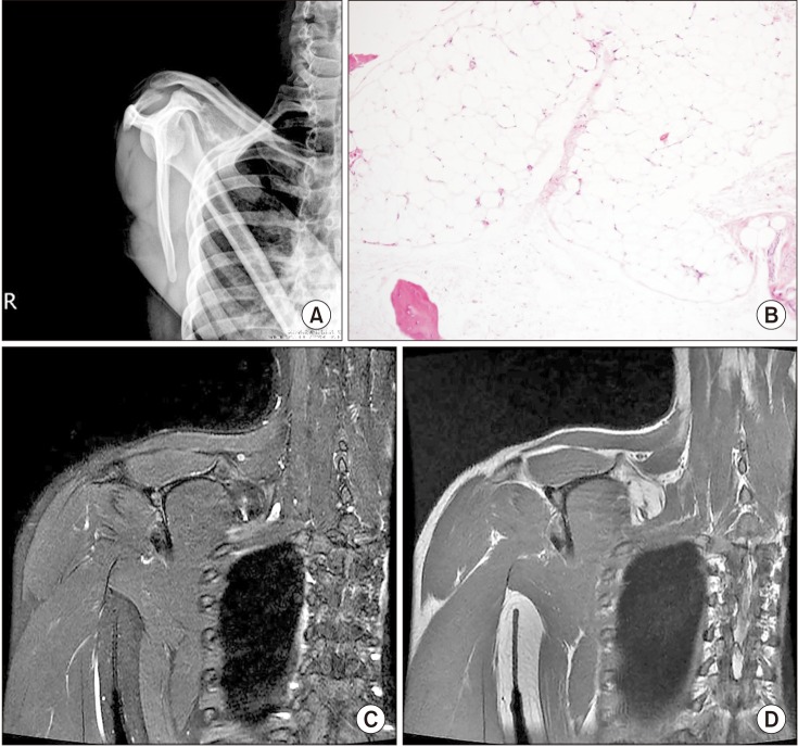
Fig. 2 A 20-year-old man with an intraosseous lipoma (early phase) in the body of scapula. (A) The outlet view shoulder radiograph demonstrates an expansile bone lesion at the body of the right scapula. (B) Photomicrograph of the lesion consisting of mature adipocytes, similar to fatty marrow. However, there is no evidence of bone marrow elements (H&E, ×100). (C) On fat-suppressed coronal T2-weighted magnetic resonance imaging, the mass shows signal dropout with fat suppression. (D) On coronal T1-weighted magnetic resonance imaging, the corresponding lesion is isointense to subcutaneous fat.
Reference
-
1. Murphey MD, Carroll JF, Flemming DJ, Pope TL, Gannon FH, Kransdorf MJ. From the archives of the AFIP: benign musculoskeletal lipomatous lesions. Radiographics. 2004; 24(5):1433–1466. PMID: 15371618.2. Milgram JW. Intraosseous lipomas: a clinicopathologic study of 66 cases. Clin Orthop Relat Res. 1988; (231):277–302.3. Shin DS, Kwak ES, Choi JH. Intraosseous lipoma. J Korean Orthop Assoc. 2003; 38(5):526–530.
Article4. Pappas AJ, Haffner KE, Mendicino SS. An intraosseous lipoma of the calcaneus: a case report. J Foot Ankle Surg. 2014; 53(5):638–642. PMID: 24875966.
Article5. Weinfeld GD, Yu GV, Good JJ. Intraosseous lipoma of the calcaneus: a review and report of four cases. J Foot Ankle Surg. 2002; 41(6):398–411. PMID: 12500792.
Article6. Campbell RS, Grainger AJ, Mangham DC, Beggs I, Teh J, Davies AM. Intraosseous lipoma: report of 35 new cases and a review of the literature. Skeletal Radiol. 2003; 32(4):209–222. PMID: 12652336.
Article7. Kim JW, Kim SJ, Kim GE, Ki SY, Lee SJ, Park JG. Radiologic findings of intraosseous lipoma of long bones. J Korean Soc Radiol. 2016; 75(4):313–321.
Article9. Bano S, Yadav SN, Chaudhary V, Jain VK. Radiological evaluation of bilateral intraosseous calcaneal lipoma in various stages of involution. Eur J Radiol Extra. 2011; 78(1):e57–e59.
Article10. Radl R, Leithner A, Machacek F, et al. Intraosseous lipoma: retrospective analysis of 29 patients. Int Orthop. 2004; 28(6):374–378. PMID: 15551133.
Article11. Muthuphei MN. Intra-osseous lipoma of the calcaneus. S Afr Med J. 1996; 86(12):1554–1555. PMID: 8998228.12. Goto T, Kojima T, Iijima T, et al. Intraosseous lipoma: a clinical study of 12 patients. J Orthop Sci. 2002; 7(2):274–280. PMID: 11956992.
Article13. Chow LT, Lee KC. Intraosseous lipoma: a clinicopathologic study of nine cases. Am J Surg Pathol. 1992; 16(4):401–410. PMID: 1566970.14. Schatz SG, Dipaola JD, D'Agostino A, Hanna R, Quinn SF. Intraosseous lipoma of the calcaneus. J Foot Surg. 1992; 31(4):381–384. PMID: 1401740.15. Greenspan A, Raiszadeh K, Riley GM, Matthews D. Intraosseous lipoma of the calcaneus. Foot Ankle Int. 1997; 18(1):53–56. PMID: 9013117.
Article16. Levin MF, Vellet AD, Munk PL, McLean CA. Intraosseous lipoma of the distal femur: MRI appearance. Skeletal Radiol. 1996; 25(1):82–84. PMID: 8717128.
Article17. Jebson PJ, Schock EJ, Biermann JS. Intraosseous lipoma of the proximal radius with extraosseous extension and a secondary posterior interosseous nerve palsy. Am J Orthop (Belle Mead NJ). 2002; 31(7):413–416. PMID: 12180628.
