Ultrasonographic and Pathologic Findings of Dermatofibrosarcoma Protuberans of the Breast: A Case Report
- Affiliations
-
- 1Department of Radiology, Dankook University Hospital, Dankook University College of Medicine, Cheonan, Korea. sirenos@hanmail.net
- 2Department of Surgery, Dankook University Hospital, Dankook University College of Medicine, Cheonan, Korea.
- 3Department of Pathology, Dankook University Hospital, Dankook University College of Medicine, Cheonan, Korea.
- KMID: 2410725
- DOI: http://doi.org/10.3348/jksr.2018.78.5.349
Abstract
- Dermatofibrosarcoma protuberans (DFSP) is a rare, malignant soft tissue tumor. It arises from the dermis, however, a few lesions are known to infiltrate into deeper layers. It commonly occurs on the trunk and extremities, but rarely involves the breast. We report a case of a 43-year-old woman who had histologically proven DFSP of the breast, with emphasis on ultrasonographic findings and pathological features. In our case, the ultrasonographic features of DFSP revealed an ovalshaped hypoechoic mass in the dermis of left breast with surrounded by increased levels of echogenic subcutaneous fat. This ultrasonographic finding correlated with pathologic features of infiltration of tumor cells into the subcutaneous fat. No previous study reported this feature of DFSP associated with the breast.
MeSH Terms
Figure
-
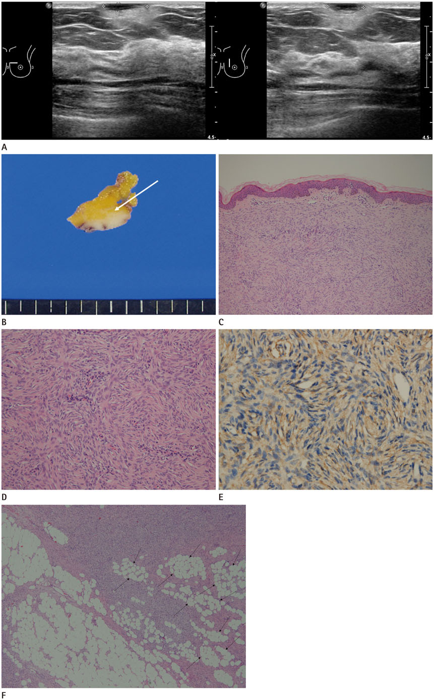
Fig. 1 A 43-year-old woman with dermatofibrosarcom protuberans, presenting with a breast mass detected on ultrasonography. A. Breast ultrasonography shows a 1.4 × 1.2 × 0.3 cm oval shape low echoic mass in the dermal layer, with surrounding increased subcutaneous fat echogenicity at the 10 o'clock direction of the left breast. B. On gross specimen, about 1.5 × 0.4 cm nodular mass locates in the dermal layer with infiltration into subcutaneous fat (arrow). C. Mammary skin shows a highly cellular spindle cell tumor proliferating just beneath the thinned epidermis (Hematoxylin and eosin stain, × 40). D. Fibroblastic tumor cells are growing closely packed and radially, in a so-called storiform pattern (Hematoxylin and eosin stain, × 200). E. The tumor cells were diffusely immunoreactive for anti-CD34 antibody, a diagnostic marker for dermatofibrosarcoma protuberans (Avidin biotin complex stain, × 400). F. The tumor cells of dermatofibrosarcoma protuberans infiltrate into deep dermis and subcutaneous fat, producing a characteristic “honeycomb pattern” (arrows) (Hematoxylin and eosin stain, × 40).
Reference
-
1. Taylor HB, Helwig EB. Dermatofibrosarcoma protuberans: a study of 115 cases. Cancer. 1962; 15:717–725.2. Mendenhall WM, Zlotecki RA, Scarborough MT. Dermatofibrosarcoma protuberans. Cancer. 2004; 101:2503–2508.
Article3. Chuang TY, Su WP, Muller SA. Incidence of cutaneous T cell lymphoma and other rare skin cancers in a defined population. J Am Acad Dermatol. 1990; 23:254–256.
Article4. Gloster HM Jr. Dermatofibrosarcoma protuberans. J Am Acad Dermatol. 1996; 35:355–374.
Article5. Fukushima H, Suda K, Matsuda M, Tanaka R, Kita H, Hanaoka T, et al. A case of dermatofibrosarcoma protuberans in the skin over the breast of a young woman. Breast Cancer. 1998; 5:407–409.
Article6. Parajuly SS, Peng YL. Sonography of dermatofibrosarcoma protuberans in the skin over breast. J Med Ultrasound. 2010; 18:130–135.
Article7. Lee SJ, Mahoney MC, Shaughnessy E. Dermatofibrosarcoma protuberance of the breast: imaging features and review of the literature. AJR Am J Roentgenol. 2009; 193:64–69.8. Kang DK, Jung YS, Yim H. Dermatofibrosarcoma protuberance of the breast: case report. J Korean Radiol Soc. 2004; 51:103–106.
Article9. Serra-Guillén C, Llombart B, Sanmartín O. [Dermatofibrosarcoma protuberans]. Actas Dermosifiliogr. 2012; 103:762–777.
Article10. Shin YR, Kim JY, Sung MS, Jung JH. Sonographic findings of dermatofibrosarcoma protuberans with pathologic correlation. J Ultrasound Med. 2008; 27:269–274.
Article
- Full Text Links
-
- Actions
-
Cited
- CITED
-
- Close
- Share
-
- Similar articles
-
- Dermatofibrosarcoma Protuberance of the Breast: Case Report
- Dermatofibrosarcoma Protuberans in Breast
- Comments to “Pigmented Dermatofibrosarcoma Protuberans Presenting as a Faint Blue Macule in a Middle-aged Korean Womanâ€
- Dermatofibrosarcoma Protuberans Arising froma Burn Scar
- A Case of Dermatofibrosarcoma Protuberans as a Subcutaneous Nodule without Surface Change
