Clinical Study of Healthy Young Men with Mitral Valve Prolapse
- Affiliations
-
- 1Department of Internal Medicine, Chosun University Medical college, Kwangju, Korea.
- 2Department of Radiology, Chosun University Medical college, Kwangju, Korea.
- KMID: 2410417
- DOI: http://doi.org/10.4250/jkse.1995.3.2.151
Abstract
- BACKGROUND
Mitral valve prolapse(MVP) is now recognized as noe of the most common cardiovascular disorders, particularly in young women with a slender body habitus. However, there is little clinical information about young men with mitral valve prolapse. The purpose of this study was to evaluate the physical and echocardiographic characteristics of young men about 20 years old with mitral valve prolapse. METHOD: Twenty young men with mitral valve prolapse(MVP graoup, mean age ; 19.9±2.4 years) and twenty healthy volunteers(control group, mean age ; 19.9±3.2 years) were examined using physical examination, chest X-ray, computed thoracic tomography and two dimensional and Doppler echocardiography.
RESULTS
Compared with control group, MVP group had a smaller chest circumference(p < 0.001), a larger height-arm span difference(p < 0.01), smaller anteroposterior chest diameters(by chest X-ray ; p < 0.001 and computed tomography ; p < 0.01) and smaller anteroposterior/transverse chest diameter ratio(chet X-ray ; p < 0.01 and computed tomography ; p < 0.05). In MVP group, mitral regurgitation was noted 15 men(75%), those had posteriorly directed jets suggesting anterior mitral leaflet anomalies.
CONCLUSION
Healthy young men having mitral valve prolapse had narrow chest and slender physical characteristics and anterior mitral leaflet abnormalities.
MeSH Terms
Figure
-
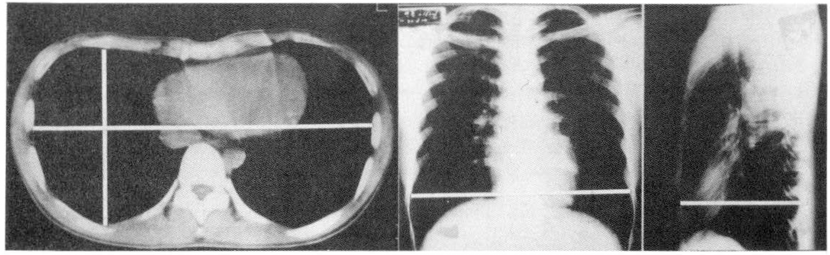
Fig. 1. Left, Computed thoracic tomography of a patient with pectus excavatum: transverse diameter measuring the inner borders of the right and left ribs at the level of the eighth thoracic vertebra, and anteroposterior diameter measuring the inner borders of the anterior and posterior ribs at the level of the eighth thoracic vertebra. Right, Posteroanterior and lateral chest X-ray films of a patient with straight back: transverse diameter measuring the inner borders of the right and left ribs at the level of the dome of diaphragm, and anteroposterior diameter measuring the inner borders of the anterior and posterior ribs at the level of the eighth thoracic vertebra.
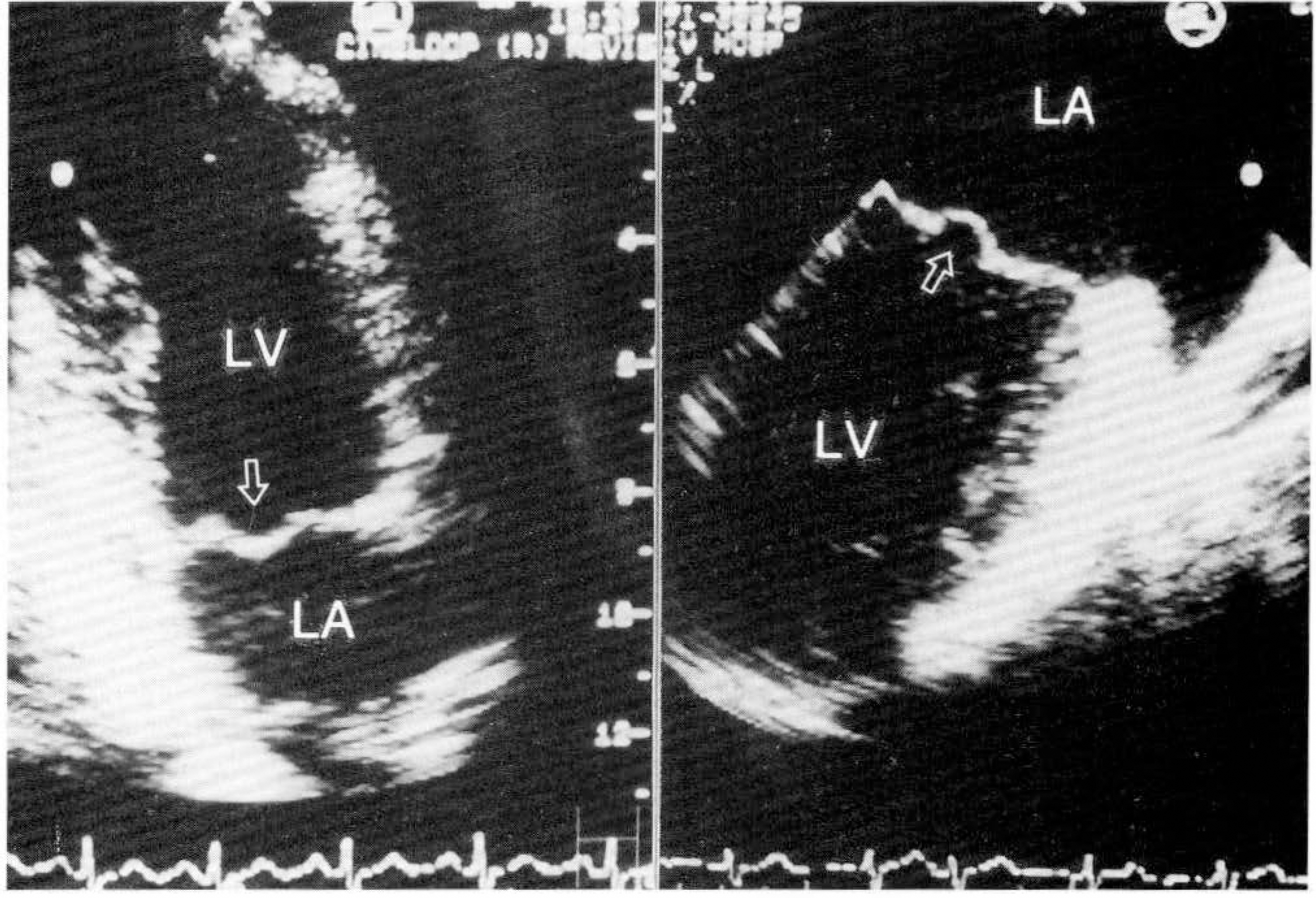
Fig. 2. Transthoracic(left) and transesophageal echocardiogram(right) show systolic doming(arrow) of the anterior mitral valve into the left atrium.
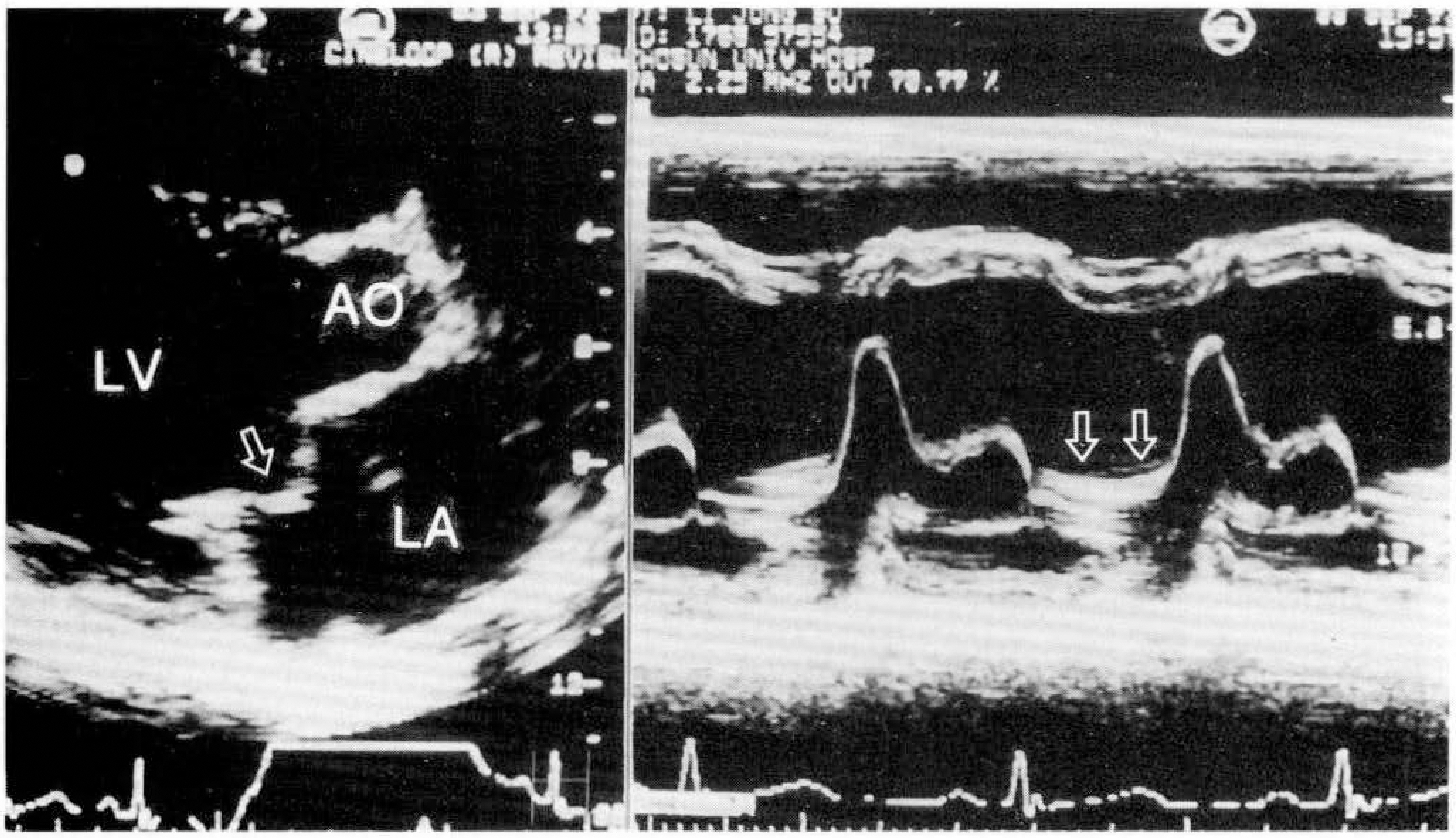
Fig. 3. Parasternal long axis echocardiograms showing systolic prolapse of the anterior mitral valve(arrow, right) and M-mode echocardiograms of the same patient showing holosystolic prolapse of the anterior mitral valve(two arrows, left).
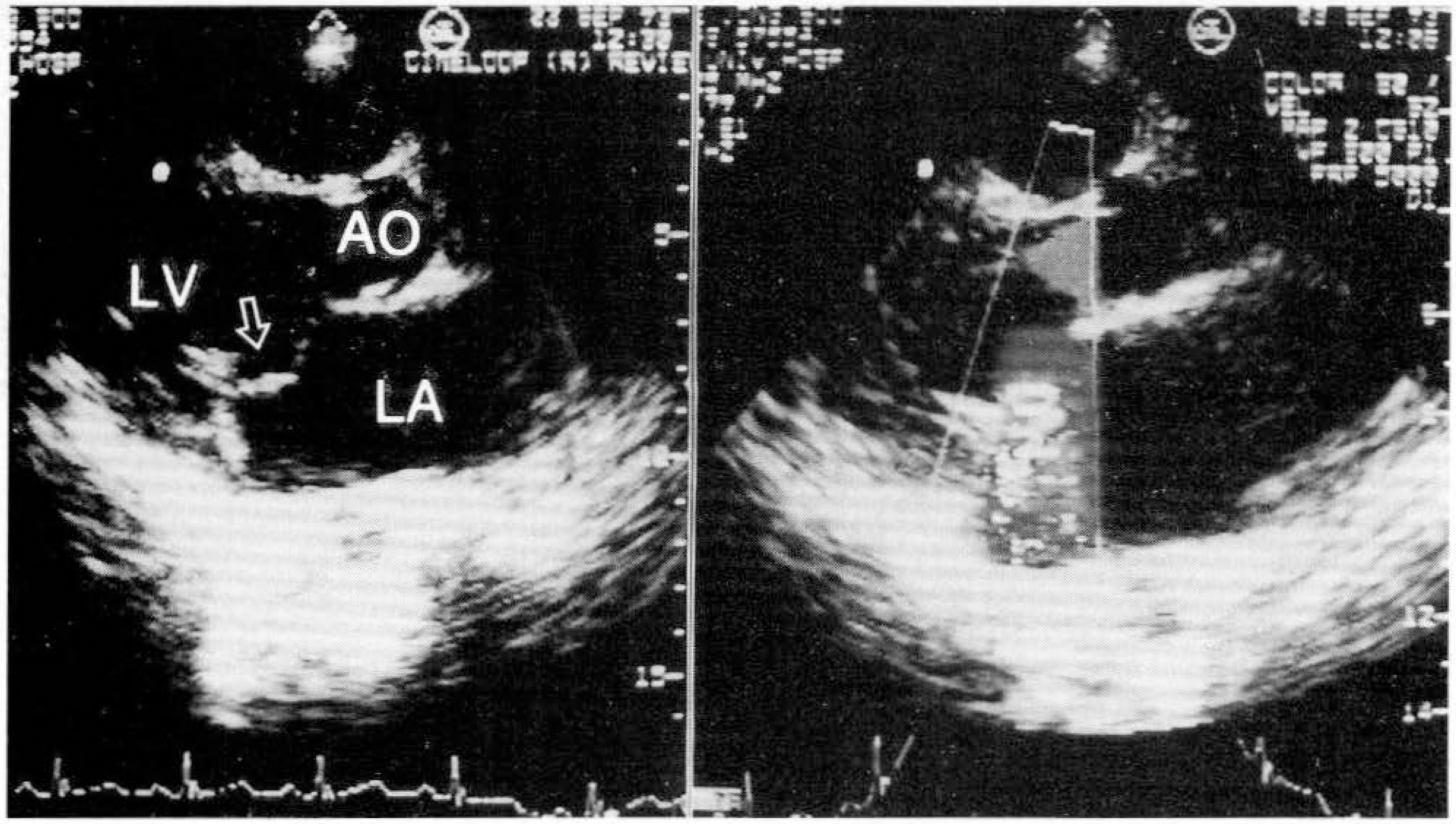
Fig. 4. Parasternal long axis views show redundancy and billowing of anterior leaflet of mitral valve(left) and posteriorly directed turbulent jet from anterior leaflet of mitral valve(right).
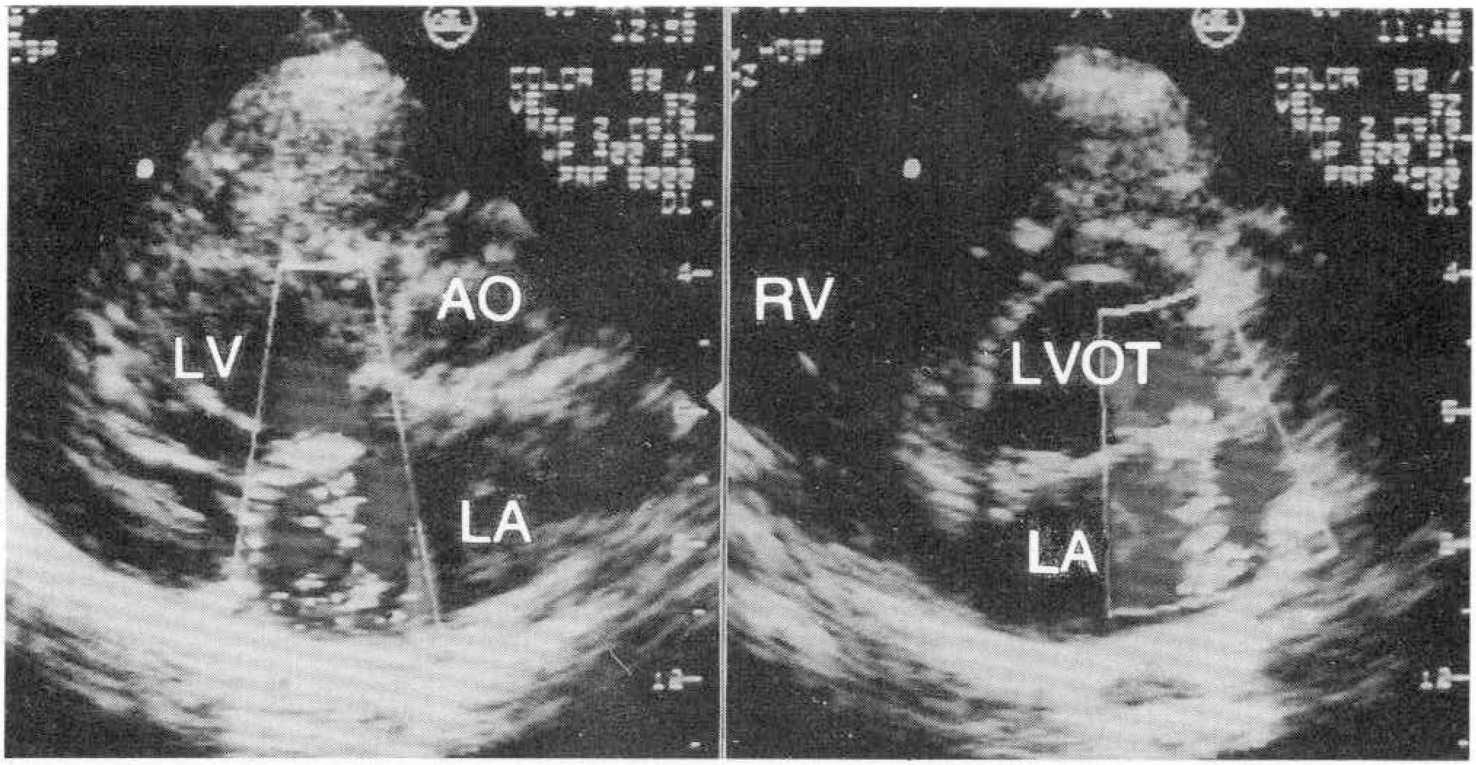
Fig. 5. Parasternal long axis view(left) shows posteriorly directed turbulent jet from anterior leaflet, and parasternal short axis view(right) shows that jet originated from lateral portion of anterior leaflet.
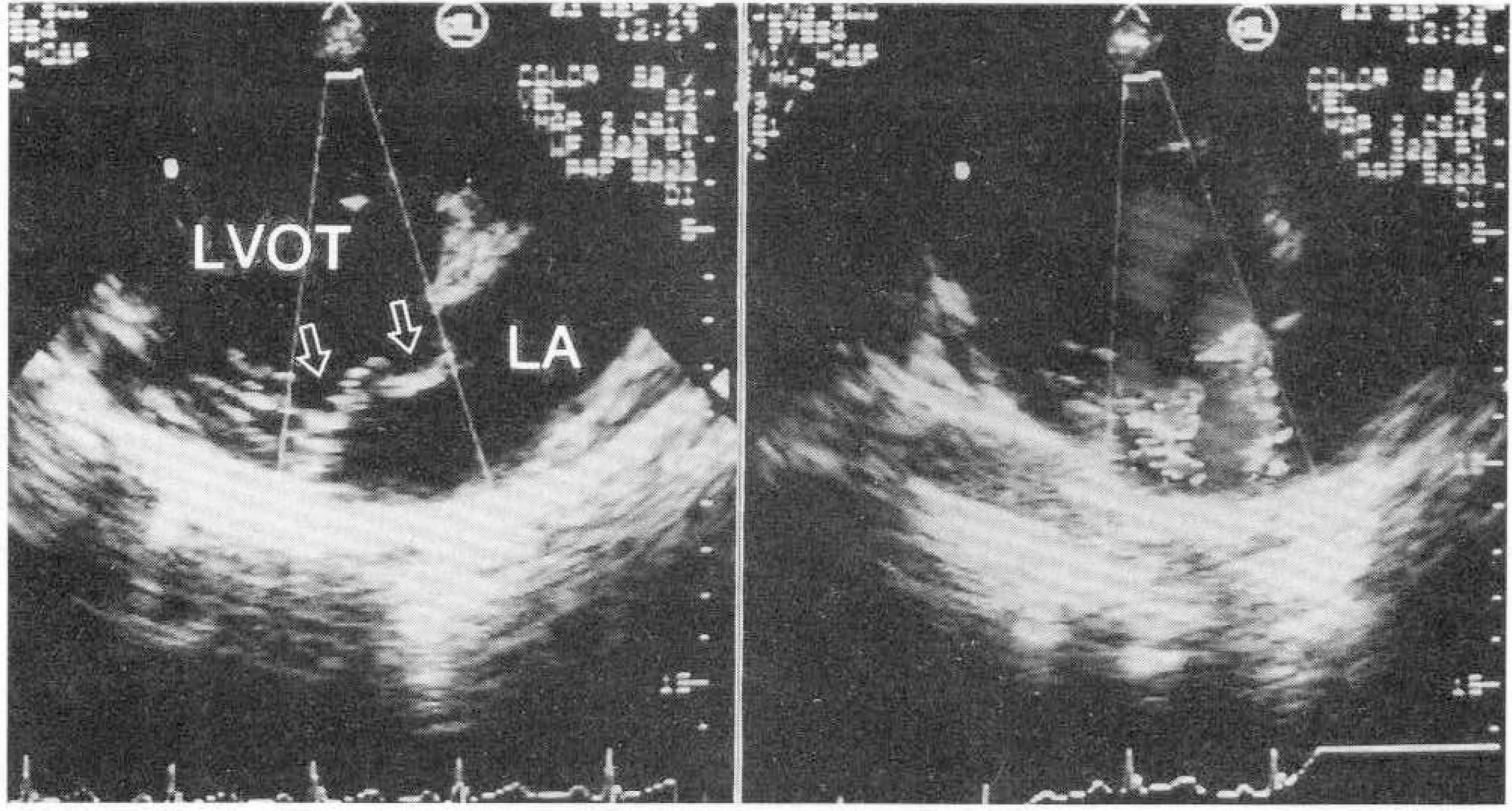
Fig. 6. Modified parasternal long axis views show redundancy of anterior leaflet of mitral valve(left, arrows) and two posteriorly directed jets from anterior leaflet of mitral valve(right).
Reference
-
References
1). Barlow JB, Pocock WA, Obel IWP. Mitral valve prolapse, primary, secondary, both or neither? Am Heart J. 102:140–146. 1981.
Article2). Devereux RB, Brown WT, Kramer-Fox R, Sachs I. Inheritance of mitral valve prolapse: effect of age and sex on gene expression. Ann Int Med. 97:826–832. 1982.
Article3). Strahan NV, Murphy EA, Fortuin NJ, Come PC, Humphries JO'N. Inheritance of the mitral valve prolapse syndrome: discussion of a three-dimensional penetrance model. Am J Med. 74:967–972. 1983.4). Fontana ME, Pence HL, Leighton RF, Wooley CF. The varying clinical spectrum of the systolic click-late systolic murmur syndrome. A postural auscultatory phenomenon. Circulation. 41:807–816. 1970.5). Epstein EJ, Coulshed N. Phonocardiogram and apex cardiogram in systolic click-late systolic murmur syndrome. Br Heart J. 35:260–275. 1973.
Article6). Devereux RB, Perloff JK, Reichek N, Josephson ME. Mitral valve prolapse. Circulation. 54:3–14. 1976.
Article7). Cheitlin MD, Sokolow M, Mcllroy MB. Clinical cardiology, sixth edition. 423. 1993.8). Bontempo CP, Ronan JA, DeLeon AC, Twigg HL. Radiographic appearance of thorax in systolic click-late systolic murmur syndrome. Am J Cardiol. 36:27–31. 1975.9). Salomon J, Shah PM, Heinle RA. Thoracic skeletal abnormalities in idiopathic mitral valve prolapse. Am J Cardiol. 36:32–36. 1975.
Article10). Schutte JE, Gaffney FA, Blend L, et al. Distinctive anthropometric characteristics of women with mitral valve prolapse. AM J Med. 71:533–538. 1981.
Article11). Boudoulas H, Wooley CF, editors. Mitral valve prolapse and the mitral valve prolapse syndrome. Mount Kisco NY: Futra Publishing Company, Inc.;1988.12). Fontana ME, Boudoulas H, Wooley CF, Sparks EA. Mitral valve prolapse and mitral valve prolapse syndrome. Curr Probl Cardiol. 311–375. 1991.13). Fontana ME. Mitral valve prolapse: physical examination and hemodynamics. Boudoulas H, Wooley CF, editors. (eds):. Mitral valve prolapse and the mitral valve prolapse syndrome. Mount Kisco. New York: Futura Publishing Company Inc;p. 217–237. 1988.14). Devereux RB. Diagnosis of mitral prolapse. Am heart J. 113:1265–1280. 1987.15). Jeresaty RM, Edwards JE, Chawla SK. Mitral valve prolapse and ruptured chordae tendineae. Am J Cardiol. 55:138–142. 1985.
Article16). Davies MJ, Moore BP, Braimbridge MV. The floppy mitral valve: Study of incidence, pathology and complications in surgical, necropsy, and forensic material. Br Heart J. 40:468–481. 1978.
Article17). Johnson LD, Caulfield JB. Morphology of myxomatous valves. Int J Cardiol. 3:373–377. 1983.18). Schaal SF. Mitral valve prolapse; Cardiac arrhythmias and electrophysiologic correlates. Boudoulas H, Wooley CF, editors. (eds):. Mitral valve prolapse and the Mitral valve prolapse syndrome. Mount Kisco. New York: Futura Publishing Company Inc;p. 567–590. 1988.19). Swartz MH, Teichholz LE, Denoso E. Mitral valve prolapse. A review of associated arrhythmias. Am J Med. 62:377–389. 1977.20). Nishimura RA, McGoon MD, Shub C, Miller FA Jr, Ilstrup DM, Tajik AJ. Echocardiographically documented mitral valve prolapse; long-term follow-up of 232 patients. N Engl J Med. 313:1305–1309. 1985.21). Beton DC, Brear SG, Edwards JD, Leonard JC. Mitral valve prolapse; an assessment of clinical features, associated conditions and prognosis. Q J Med. 52:150–164. 1983.22). Devereux RB, Kramer-Fox R, Brown WT, et al. Relation between clinical features of the mitral prolapse syndrome and echocardiographically documented mitral valve prolapse. J Am Coll Cardiol. 8:763–772. 1986.
Article23). Ranganathan N, Silver MD, Robinson TI, et al. Angiographic-morphologic correlation in patients with severe mitral regurgitation due to prolapse of the posterior mitral valve leaflet. Circulation. 48:514–518. 1973.
Article
- Full Text Links
-
- Actions
-
Cited
- CITED
-
- Close
- Share
-
- Similar articles
-
- Combined Mitral and Aortic Valve Prolapse
- Echocardiographic Analysis of Systolic Mitral Valve Motion in Healthy Young Males: With Particular Reference to Mitral Valve Prolapse
- Congenital Double-Orifice Mitral Valve with Mitral Valve Prolapse and Severe Mitral Regurgitation
- Mitral Valve Prolapse in Patients with Panic Attacks in Korea
- Mitral valve prolapse
