J Korean Soc Echocardiogr.
1995 Dec;3(2):115-120. 10.4250/jkse.1995.3.2.115.
Echocardiographic Detection of Left Ventricular Asynergy by Apical Rotational Transducer Approach in Ischemic Heart Disease
- Affiliations
-
- 1Department of Internal Medicine, Wonkwang University, School of Medicine, Iksan, Korea.
- KMID: 2410412
- DOI: http://doi.org/10.4250/jkse.1995.3.2.115
Abstract
OBJECTIVE
To compare the usefulness of the apical transducer approach with that of parasternal approach in two-dimensional echocardiography for detection of left ventricular(LV) regional asynergy in ischemic heart diesease, the 49 subjects(25 of acute myocardial infarction, 7 of old myocardial infarction with angian, 16 of unstable angina pectoris) having good echocardiographic window with significant coronary lesion were studied.
METHODS
We assessed the LV regional asynergy by visual analysis and scoring of 16 LV wall segments(8 in proximal, 8 in distal) from video playback. We compared the above two approaches using the same scoring scale recommended by American Society of Echocardiography.
RESULTS
1) All 6 patients with old myocardial infarction, 20(80%) of 25 patients with acute myocardial infarction, 6(33%) of 18 patients with unstable angina showed apparent LV asynergy on either approach. 2) There was a overall good agreement(89%, 131 of 147 segments) between two approaches in detecting LV regional asynergy when dividing the LV into 3 regions(anterior, lateral, posterior). 3) Of the total 16 disagreed regions, 13(81%) regions detected only by the apical approach were localized to apex of th LV, and 3(19%) regions detected only by the parasternal approach were mostly localized to base of the LV. 4) Of toal 19 dyskinetic segments, 3(16%) segments were detected by the parasternal approach, while 16(84%) segments by the apical approach. 5) All 7 aneurysmal segments were detected only by the apical approach.
CONCLUSION
Both the apical and the parasternal approaches are complementary each other, but the apical approach might be a simple, reliable method providing better images for detection of LV regional asynergy, which localized near the cardiac apex and compares favorably with the parasternal short axis approach.
MeSH Terms
Figure
-

Fig. 1. Echocardiographic Assessment of Regional Wall Motion with Modified 16-segment Model. Ant.=anterior wall; Lat. = lateral wall; Post.=posterior wall; Apical=apical rotational approach.
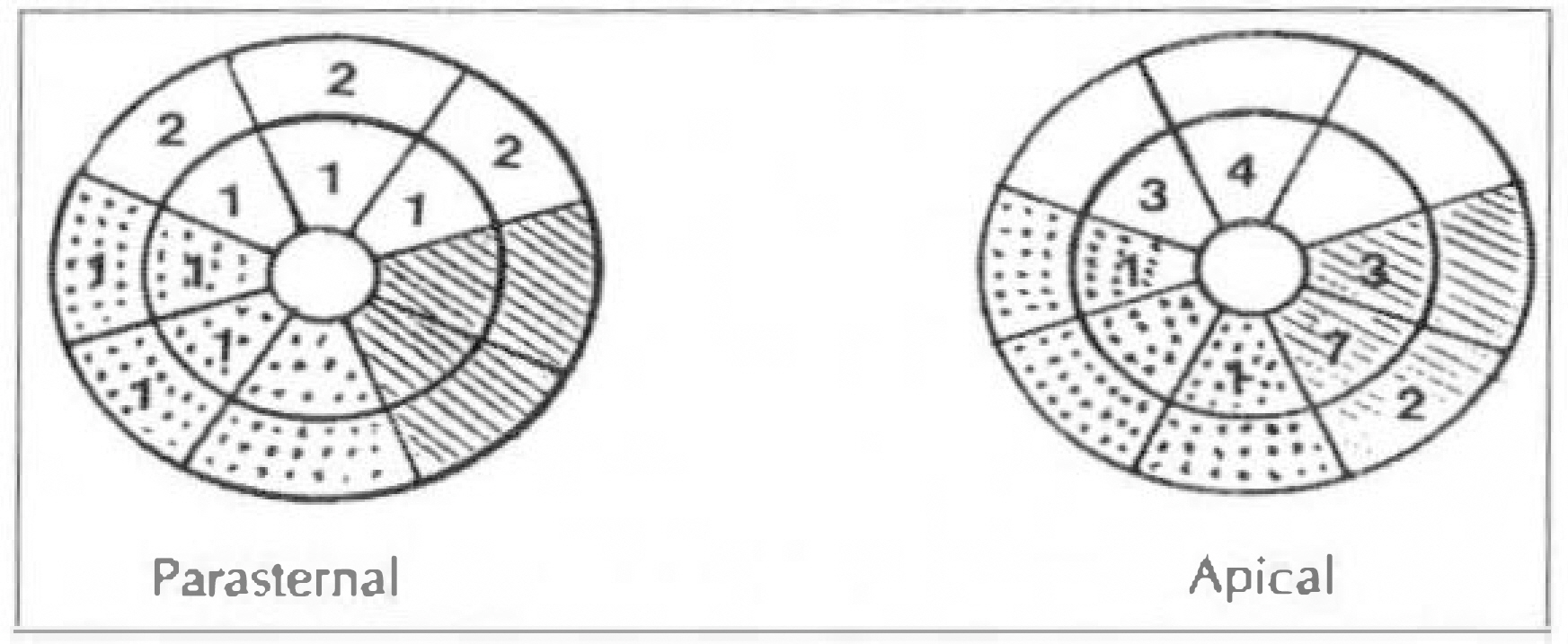
Fig. 2. Schematic Bull Eye Diagram Showing Number of Disagreed Segments in Each Method of Approach. clean sector=anterior wall; hatched sector=lateral wall; dotted sector=posterior wall.
Reference
-
References
1). Parisi AF, Moynihan PF, Folland ED, Feldman CL. Quantitative detection of regional left ventricular contraction abnormalities by two dimensional echocardiography. II. Accuracy in coronary artery disease. Circulation. 63:761. 1981.2). Zoghbi WA, Charlat ML, Bolli R, Zhu WX, Hartley CJ, Quinones MA. Quantitative assessment of left ventricular wall motion by two dimensional echocardiography: Validation during reversible ischemia in the conscious dog. J Am Coll Cardiol. 11:851. 1988.3). Lundgren C, Bourdillon PDV, Dillon JC, Feigenbaum H. Comparison of contrast angiography and two-dimensional echocardiography for the evaluation of left ventricular regional wall motion abnormalities after acute myocardial infarction. Am J Cardiol. 65:1071. 1990.
Article4). Henschke CI, Risser TA, Sandor T, Hanlon WB, Neumann A, Wynne J. Quantitative computer-assisted analysis of left ventricular wall thickening and motion by 2-dimensional echocardiography in acute myocardial infarction. Am J Cardiol. 52:960. 1983.
Article5). Horowitz RS, Morganroth J, Parrotto C, Chen CC, Soffer J, Pauletto FJ. Immediate diagnosis of acute myocardial infarction by two-dimensional echocardiography. Circulation. 65:323. 1982.
Article6). Sheehan FH, Stewart DK, Dodge HT, Mitten S, Bolson EL, Brown G. Variability in the measurement of regional left ventricular wall motion from contrast angiograms. Circulation. 68:550. 1983.
Article7). Erbel R, Schweizer P, Lambertz H, Henn G, Mryer J, Krebs W, Effert S. Echoventriculography-A simultaneous analysis of two-dimensional echocardiography and cineventriculography. Circulation. 67:205. 1983.8). Kisslo JA, Robertson D, Ritjerford JD, Gilbert BW, Romm OV, Behar VS. A comparison of real-time, two-dimensional echocardiography and cineangiography in detecting left ventricular asynergy. Circulation. 55:134. 1977.9). Guyer DE, Goale RA, Gillam LD, Wilkins GT, Guerrero JL, Weyman AE. An echocardiographic technique for quantifying and displaying the extent of regional left ventricular dyssynergy. J Am Coll Cardol. 8:830. 1986.
Article10). Schnittger I, Fitzgerald PJ, Gordon EP, Alderman EL, Popp RL. Computerized quantitative analysis of left ventricular wall motion by two dimensional echocardiography. Circulation. 70:242. 1984.11). Feigenbaum H. Assessing the left ventricle with two-dimensional echocardiography. 2:305. 1989.12). Schiller NB, Shah PM, Crawford M, DeMaria A, et al. Recommendations for quantitation of the left ventricle by two-dimensional echocardiography. J Am Soc Echo. 2:358. 1989.
Article13). Ginzton LE, Conant R, Brizendine M, Lee F, Mena I, Laks MM. Exercise subcostal two-dimensional echocardiography: A new method of segmental wall motion analysis. Am J Cardiol. 53:805. 1984.
Article14). Crouse LJ, Harbrecht JJ, Vacek JL, Rosamond TL, Kramer PH. Exercise echocardiography as a screening test for coronary artery disease and correlation with coronary arteriography. Am J Cardiol. 67:1213. 1991.
Article15). Limacher MC, Quinones MA, Poliner LR, Nelson JG, Winters WL, Waggoner AD. Detection of coronary artery disease with exercise two-dimensional echocardiography. Circulation. 67:1211. 1983.16). Assmann P, Stager CJ, Borden SG, Dreysse ST, Tijssen JP, Sutherland GR, Roelandt JR. Quantitative echocardiographic analysis of global and regional left ventricular function: A problem revisited. J Am Soc Echo. 3:478. 1990.
Article17). Assmann P, Stager CJ, Borden SG, Sutherland GR, Roelandt JR. Reference systems in echocardiographic quantitative wall motion analysis with registration of respiration. J Am Soc Echo. 4:224. 1991.
Article18). Bourdillon PDV, Broderick TM, Sawada SG, Armstrong WF, Ryan T, Dillon JC, Fineberg NS, Feigenbaum H. Regional wall motion index for infarct and noninfarct regions after reperfusion in acute myocardial infarction: Comparison with global wall motion index. J Am Soc Echo. 2:398. 1989.
Article
- Full Text Links
-
- Actions
-
Cited
- CITED
-
- Close
- Share
-
- Similar articles
-
- Multiple Coronary Artery-Left Ventricular Microfistulae in a Patient with Apical Hypertrophic Cardiomyopathy: A Demonstration by Transthoracic Color Doppler Echocardiography
- A Novel Cardiomyopathy Mimicking Acute Myocardial Infarction
- Surgery for a Muscular Type Ventricular Septal Defect via Right Apical Ventriculotomy: A case report
- Real Time Determination of Left Atrial Volume by Automatic Boundary Detection: Comparison with Two-dimensional Echocardiographic Measurement
- The Heart in Acute Glomerulonephritis: An Echocardiographic Study
