J Korean Orthop Assoc.
2018 Apr;53(2):121-128. 10.4055/jkoa.2018.53.2.121.
Prognostic Factors in Acetabular Development Following Reduction of Developmental Dislocation of the Hip in Patients under the Age of 24 Months
- Affiliations
-
- 1Department of Orthopaedic Surgery, Chonnam National University Hospital, Gwangju, Korea. stjung@chonnam.ac.kr
- KMID: 2410059
- DOI: http://doi.org/10.4055/jkoa.2018.53.2.121
Abstract
- PURPOSE
The aims of this study are to evaluate the outcome of treatment for developmental dislocation of the hip (DDH) in children under the age of 24 months who underwent open reduction (OR) or closed reduction (CR) and to determine radiologic prognostic factor.
MATERIALS AND METHODS
A total of 90 hips of 88 children under the age of 24 months treated for DDH were included. The treatments for these children were CR in 29 hips and OR in 61 hips. All patients were followed up for more than 5 years. Radiographic evaluations, including acetabular index (AI), Yamamuro's distance a and b, center-edge angle (CEA), sourcil shape, and teardrop shape have been proposed to indicate the degree of DDH. Hips were reclassified according to the Severin criteria (classes I and II, satisfactory; classes III and IV, unsatisfactory).
RESULTS
Among the 90 hips, 67 hips (74.4%) were included in the "˜satisfactory group', while 23 hips (25.6%) were included in the "˜unsatisfactory group'. In the CR group, 23 hips (79.3%) were included in the "˜satisfactory group', while 6 hips (20.7%) were included in the "˜unsatisfactory group'. In the OR group, 44 hips (72.1%) were included in the "˜satisfactory group', while 17 hips (27.9%) were included in the "˜unsatisfactory group'. There was no significant difference between the reduction methods. At 1 year follow-up after reduction, the AI improvement in the "˜satisfactory group' (8.1° [23.4%]) was significantly higher than that in the "˜unsatisfactory group' (6.7° [18.5%]) (p=0.012). A significant difference of the mean CEA values was observed between the "˜satisfactory group' and the "˜unsatisfactory group' 3 years after the treatment (p=0.001). Five years after reduction, the V shape of teardrop and the upward shape of acetabular sourcil were observed in 2 hips (3.0%) and 4 hips (6.0%) of the "˜satisfactory group', respectively, whereas the corresponding findings were observed in 3 hips (13.0%) and 5 hips (21.7%) of the "˜unsatisfactory group', respectively (p=0.023, 0.005).
CONCLUSION
The improvement of AI at 1-year and CEA at 3-year follow-ups, as well as teardrop shape and sourcil shape at 5-year followup, were reliable radiographic prognostic factor of DDH.
Figure
-
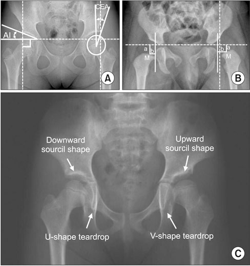
Figure 1 Radiologic measurements used for predicting acetabular development after surgery. (A) Acetabular index (AI) and center-edge angle (CEA). (B) Yamamuro's distance a & b. (C) Sourcil shape and teardrop shape. M, center of metaphysis.

Figure 2 Changes in the acetabular index during a follow-up of patients with DDH between the CR and OR groups. *By Student t-test. DDH, developmental dislocation of the hip, CR, closed reduction; OR, open reduction; POD, postoperative day.

Figure 3 Comparison of changes in the acetabular index between the satisfactory and unsatisfactory groups. *By Student t-test. POD, postoperative day.
Reference
-
1. Gholve PA, Flynn JM, Garner MR, Millis MB, Kim YJ. Predictors for secondary procedures in walking DDH. J Pediatr Orthop. 2012; 32:282–289.
Article2. Morel G. The treatment of congenital dislocation and subluxation of the hip in the older child. Acta Orthop Scand. 1975; 46:364–399.3. Rampal V, Sabourin M, Erdeneshoo E, Koureas G, Seringe R, Wicart P. Closed reduction with traction for developmental dysplasia of the hip in children aged between one and five years. J Bone Joint Surg Br. 2008; 90:858–863.
Article4. Bulut M, Gürger M, Belhan O, Batur OC, Celik S, Karakurt L. Management of developmental dysplasia of the hip in less than 24 months old children. Indian J Orthop. 2013; 47:578–584.
Article5. Farsetti P, Caterini R, Potenza V, Ippolito E. Developmental dislocation of the hip successfully treated by preoperative traction and medial open reduction: a 22-year mean followup. Clin Orthop Relat Res. 2015; 473:2658–2669.
Article6. Castillo R, Sherman FC. Medial adductor open reduction for congenital dislocation of the hip. J Pediatr Orthop. 1990; 10:335–340.
Article7. Mergen E, Adyaman S, Omeroglu H, Erdemli B, Isiklar U. Predictors for secondary procedures in walking DDH. J Pediatr Orthop. 2012; 32:282–289.8. Harris NH. Acetabular growth potential in congenital dislocation of the hip and some factors upon which it may depend. Clin Orthop Relat Res. 1976; 119:99–106.
Article9. Li Y, Xu H, Li J, et al. Early predictors of acetabular growth after closed reduction in late detected developmental dysplasia of the hip. J Pediatr Orthop B. 2015; 24:35–39.
Article10. Ward WT, Vogt M, Grudziak JS, Tümer Y, Cook PC, Fitch RD. Severin classification system for evaluation of the results of operative treatment of congenital dislocation of the hip. A study of intraobserver and interobserver reliability. J Bone Joint Surg Am. 1997; 79:656–663.
Article11. Roposch A, Wedge JH, Riedl G. Reliability of Bucholz and Ogden classification for osteonecrosis secondary to developmental dysplasia of the hip. Clin Orthop Relat Res. 2012; 470:3499–3505.
Article12. Boniforti FG, Fujii G, Angliss RD, Benson MK. The reliability of measurements of pelvic radiographs in infants. J Bone Joint Surg Br. 1997; 79:570–575.
Article13. Yamamuro T, Chene SH. A radiological study on the development of the hip joint in normal infants. J Jpn Orthop Assoc. 1975; 49:421–439.14. Kahle WK, Coleman SS. The value of the acetabular teardrop figure in assessing pediatric hip disorders. J Pediatr Orthop. 1992; 12:586–591.
Article15. Kim HT, Kim JI, Yoo CI. Diagnosing childhood acetabular dysplasia using the lateral margin of the sourcil. J Pediatr Orthop. 2000; 20:709–717.
Article16. Brougham DI, Broughton NS, Cole WG, Menelaus MB. The predictability of acetabular development after closed reduction for congenital dislocation of the hip. J Bone Joint Surg Br. 1988; 70:733–736.
Article17. Race C, Herring JA. Congenital dislocation of the hip: an evaluation of closed reduction. J Pediatr Orthop. 1983; 3:166–172.18. Gibson PH, Benson MK. Congenital dislocation of the hip. Review at maturity of 147 hips treated by excision of the limbus and derotation osteotomy. J Bone Joint Surg Br. 1982; 64:169–175.
Article19. Powell EN, Gerratana FJ, Gage JR. Open reduction for congenital hip dislocation: the risk of avascular necrosis with three different approaches. J Pediatr Orthop. 1986; 6:127–132.20. Albinana J, Dolan LA, Spratt KF, Morcuende J, Meyer MD, Weinstein SL. Acetabular dysplasia after treatment for developmental dysplasia of the hip. Implications for secondary procedures. J Bone Joint Surg Br. 2004; 86:876–886.21. Zamzam MM, Kremli MK, Khoshhal KI, et al. Acetabular cartilaginous angle: a new method for predicting acetabular development in developmental dysplasia of the hip in children between 2 and 18 months of age. J Pediatr Orthop. 2008; 28:518–523.22. Shin CH, Yoo WJ, Park MS, Kim JH, Choi IH, Cho TJ. Acetabular remodeling and role of osteotomy after closed reduction of developmental dysplasia of the hip. J Bone Joint Surg Am. 2016; 98:952–957.
Article23. Kitoh H, Kitakoji T, Katoh M, Ishiguro N. Prediction of acetabular development after closed reduction by overhead traction in developmental dysplasia of the hip. J Orthop Sci. 2006; 11:473–477.
Article24. Gotoh E, Tsuji M, Matsuno T, Ando M. Acetabular development after reduction in developmental dislocation of the hip. Clin Orthop Relat Res. 2000; 378:174–182.
Article25. Albiñana J, Morcuende JA, Delgado E, Weinstein SL. Radiologic pelvic asymmetry in unilateral late-diagnosed developmental dysplasia of the hip. J Pediatr Orthop. 1995; 15:753–762.
Article26. Zionts LE, MacEwen GD. Treatment of congenital dislocation of the hip in children between the ages of one and three years. J Bone Joint Surg Am. 1986; 68:829–846.
Article27. Malvitz TA, Weinstein SL. Closed reduction for congenital dysplasia of the hip. Functional and radiographic results after an average of thirty years. J Bone Joint Surg Am. 1994; 76:1777–1792.
Article28. Novais EN, Hill MK, Carry PM, Heyn PC. Is age or surgical approach associated with osteonecrosis in patients with developmental dysplasia of the hip? A meta-analysis. Clin Orthop Relat Res. 2016; 474:1166–1177.
Article29. Gardner RO, Bradley CS, Howard A, Narayanan UG, Wedge JH, Kelley SP. The incidence of avascular necrosis and the radiographic outcome following medial open reduction in children with developmental dysplasia of the hip: a systematic review. Bone Joint J. 2014; 96:279–286.
- Full Text Links
-
- Actions
-
Cited
- CITED
-
- Close
- Share
-
- Similar articles
-
- Arthrographic Evaluation in Developmental Dislocation of the Hip: Comparison Between Hip Arthrogram and Operative Findings
- Hip Development after Reduction in Developmental Dislocation of the Hip: Long-term Follow-up to Skeletal Maturity of 64 Hips
- Avasular Nevrosis and Acetabular Dysplasia Following Treatment of Developmental Dislocation of the Hip
- Predicatable Factors of Developmental Dislocation of the Hip after Reduction
- Acetabular Dysplasia and Osteoarthritis Developed by an Eversion of the Acetabular Labrum
