Subtalar Arthroscopy and Posterior Endoscopy
- Affiliations
-
- 1Department of Orthopaedic Surgery, Seoul St. Mary's Hospital, College of Medicine, The Catholic University of Korea, Seoul, Korea. jahn@catholic.ac.kr
- KMID: 2410056
- DOI: http://doi.org/10.4055/jkoa.2018.53.2.93
Abstract
- The application of arthroscopy is becoming increasingly widespread due to the development of surgical instruments and techniques. Subtalar pathology can cause chronic pain in the hindfoot, but it is often misdiagnosed as a lesion of the adjacent ankle joint, which can lead to delayed diagnosis and treatment. Subtalar arthroscopy and posterior endoscopy are good methods to confirm and treat the posterior pathology of the subtalar joint and posterior ankle joint.
MeSH Terms
Figure
-
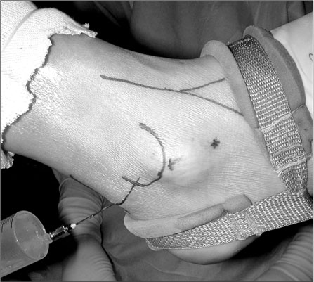
Figure 1 Photograph showing the three portals used in subtalar arthroscopy. Normal saline is injected into the subtalar joint through the posterolateral portal.
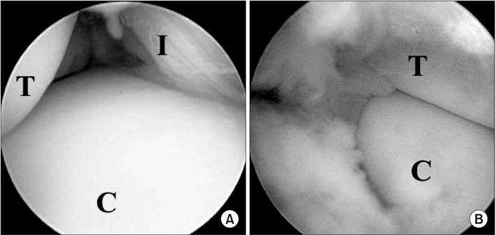
Figure 2 Arthroscopic findings showing the inside view of the subtalar joint. (A) Zone 1, which is the anteromedial side of posterior calcaneal facet, is viewed through the middle portal. (B) Zone 4, which is the anterolateral side of subtalar joint, is viewed through the anterolateral portal. T, talus; C, calcaneus; I, interosseous talocalcaneal ligament.
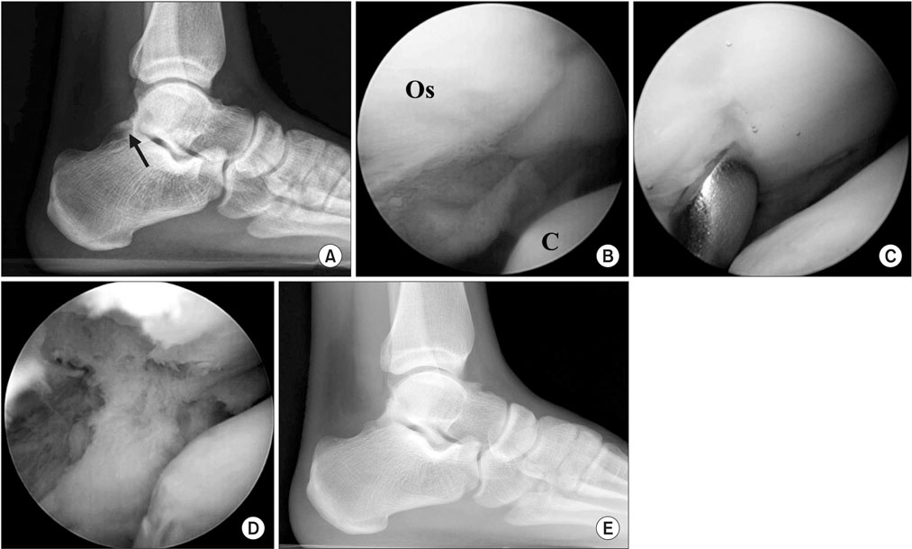
Figure 3 (A) Standing lateral radiograph of the right foot of a 27-year-old male patient showing painful Os trigonum (Os) (arrow). (B) This arthroscopic finding demonstrates large Os. C, calcaneus. (C) The Os is mobilized with an arthroscopic curette. (D) The Os is removed completely. (E) Standing lateral radiograph at postoperative 1 year shows the cleared posterior ankle space with removal of Os.

Figure 4 Photograph shows the posterior two-portal posterior endoscopic procedure.
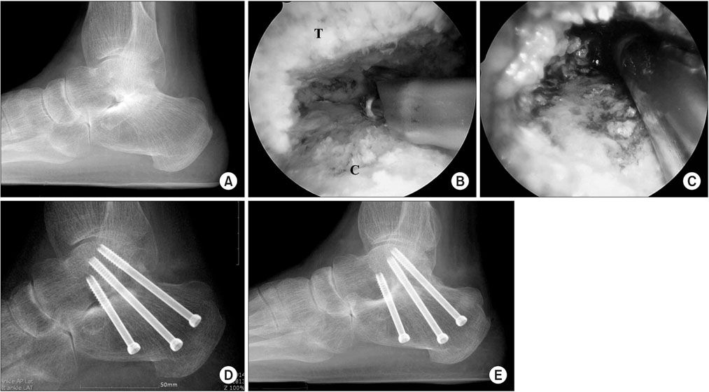
Figure 5 (A) Standing lateral radiograph of the left foot of a 52-year-old male rheumatoid arthritis patient showing severe narrowing of the subtalar joint space. (B) Posterior endoscopic finding shows the preparation of joint surface with a burr. T, talus; C, calcaneus. (C) Morsellized bone grafts are inserted through the arthroscopic sheath. (D) Lateral radiograph taken 2 weeks postoperatively shows that the subtalar joint is well fixated with 3 screws. (E) Radiograph taken 14 months postoperatively demonstrates satisfactorily fused subtalat joint.
Reference
-
1. Ahn JH, Choy WS, Lee KW. Arthroscopy of the first metatarsophalangeal joint in 59 consecutive cases. J Foot Ankle Surg. 2012; 51:161–167.
Article2. Kuyucu E, Mutlu H, Mutlu S, Gülenç B, Erdil M. Arthroscopic treatment of focal osteochondral lesions of the first metatarsophalangeal joint. J Orthop Surg Res. 2017; 12:95.
Article3. Parisien JS, Vangsness T. Arthroscopy of the subtalar joint: an experimental approach. Arthroscopy. 1985; 1:53–57.4. Muñoz G, Eckholt S. Subtalar arthroscopy: indications, technique and results. Foot Ankle Clin. 2015; 20:93–108.5. Lui TH, Tong SC. Subtalar arthroscopy: when, why and how. World J Orthop. 2015; 6:56–61.
Article6. Williams MM, Ferkel RD. Subtalar arthroscopy: indications, technique, and results. Arthroscopy. 1998; 14:373–381.
Article7. Frey C, Gasser S, Feder K. Arthroscopy of the subtalar joint. Foot Ankle Int. 1994; 15:424–428.
Article8. van Dijk CN, Scholten PE, Krips R. A 2-portal endoscopic approach for diagnosis and treatment of posterior ankle pathology. Arthroscopy. 2000; 16:871–876.
Article9. Ogut T, Ayhan E, Irgit K, Sarikaya AI. Endoscopic treatment of posterior ankle pain. Knee Surg Sports Traumatol Arthrosc. 2011; 19:1355–1361.
Article10. Frey C. Surgical advancements: arthroscopic alternatives to open procedures: great toe, subtalar joint, Haglund's deformity, and tendoscopy. Foot Ankle Clin. 2009; 14:313–339.
Article11. van Dijk CN. Hindfoot endoscopy. Foot Ankle Clin. 2006; 11:391–414. vii
Article12. Viladot A, Lorenzo JC, Salazar J, Rodríguez A. The subtalar joint: embryology and morphology. Foot Ankle. 1984; 5:54–66.
Article13. Stephens MM, Sammarco GJ. The stabilizing role of the lateral ligament complex around the ankle and subtalar joints. Foot Ankle. 1992; 13:130–136.
Article14. Harper MC. The lateral ligamentous support of the subtalar joint. Foot Ankle. 1991; 11:354–358.
Article15. Rammelt S, Gavlik JM, Barthel S, Zwipp H. The value of subtalar arthroscopy in the management of intra-articular calcaneus fractures. Foot Ankle Int. 2002; 23:906–916.
Article16. Tasto JP. Arthroscopy of the subtalar joint and arthroscopic subtalar arthrodesis. Instr Course Lect. 2006; 55:555–564.17. Elgafy H, Ebraheim NA. Subtalar arthroscopy for persistent subfibular pain after calcaneal fractures. Foot Ankle Int. 1999; 20:422–427.
Article18. Lee KB, Saltzman CL, Suh JS, Wasserman L, Amendola A. A posterior 3-portal arthroscopic approach for isolated subtalar arthrodesis. Arthroscopy. 2008; 24:1306–1310.
Article19. Jerosch J. Subtalar arthroscopy: indications and surgical technique. Knee Surg Sports Traumatol Arthrosc. 1998; 6:122–128.20. de Leeuw PA, Golanó P, Sierevelt IN, van Dijk CN. The course of the superficial peroneal nerve in relation to the ankle position: anatomical study with ankle arthroscopic implications. Knee Surg Sports Traumatol Arthrosc. 2010; 18:612–617.
Article21. Tryfonidis M, Whitfield CG, Charalambous CP, Baraza WK, Blundell C, Sharp RJ. The distance between the sural nerve and ideal portal placements in lateral subtalar arthroscopy: a cadaveric study. Foot Ankle Int. 2008; 29:842–844.
Article22. Mekhail AO, Heck BE, Ebraheim NA, Jackson WT. Arthroscopy of the subtalar joint: establishing a medial portal. Foot Ankle Int. 1995; 16:427–432.
Article23. Ferkel RD. Foot and ankle arthroscopy. 2nd ed. Philadelphia: Wolters Kluwer;2017. p. 335–352.24. Frey C, Feder KS, DiGiovanni C. Arthroscopic evaluation of the subtalar joint: does sinus tarsi syndrome exist? Foot Ankle Int. 1999; 20:185–191.
Article25. Marumoto JM, Ferkel RD. Arthroscopic excision of the os trigonum: a new technique with preliminary clinical results. Foot Ankle Int. 1997; 18:777–784.
Article26. Ahn JH, Lee SK, Kim KJ, Kim YI, Choy WS. Subtalar arthroscopic procedures for the treatment of subtalar pathologic conditions: 115 consecutive cases. Orthopedics. 2009; 32:891.
Article27. Lee KB, Bai LB, Song EK, Jung ST, Kong IK. Subtalar arthroscopy for sinus Tarsi syndrome: arthroscopic findings and clinical outcomes of 33 consecutive cases. Arthroscopy. 2008; 24:1130–1134.
Article28. Sitte W, Lampert C, Baumann P. Osteosynthesis of talar body shear fractures assisted by hindfoot and subtalar arthroscopy: technique tip. Foot Ankle Int. 2012; 33:74–78.
Article29. Martin DF, Baker CL, Curl WW, Andrews JR, Robie DB, Haas AF. Operative ankle arthroscopy. Long-term followup. Am J Sports Med. 1989; 17:16–23. discussion 23.30. Ferkel RD, Small HN, Gittins JE. Complications in foot and ankle arthroscopy. Clin Orthop Relat Res. 2001; 391:89–104.
Article31. Nault ML, Kocher MS, Micheli LJ. Os trigonum syndrome. J Am Acad Orthop Surg. 2014; 22:545–553.
Article32. Guo QW, Hu YL, Jiao C, Yu CL, Ao YF. Arthroscopic treatment for osteochondral lesions of the talus: analysis of outcome predictors. Chin Med J (Engl). 2010; 123:296–300.33. Zengerink M, Struijs PA, Tol JL, van Dijk CN. Treatment of osteochondral lesions of the talus: a systematic review. Knee Surg Sports Traumatol Arthrosc. 2010; 18:238–246.
Article34. Saxena A, Perez H. Pigmented villonodular synovitis about the ankle: a review of the literature and presentation in 10 athletic patients. Foot Ankle Int. 2004; 25:819–826.
Article35. Doral MN, Uzumcugil A, Bozkurt M, et al. Arthroscopic treatment of synovial chondromatosis of the ankle. J Foot Ankle Surg. 2007; 46:192–195.
Article36. Ogut T, Ayhan E. Hindfoot endoscopy for accessory flexor digitorum longus and flexor hallucis longus tenosynovitis. Foot Ankle Surg. 2011; 17:e7–e9.
Article37. Grant TH, Kelikian AS, Jereb SE, McCarthy RJ. Ultrasound diagnosis of peroneal tendon tears. A surgical correlation. J Bone Joint Surg Am. 2005; 87:1788–1794.38. de Leeuw PA, van Sterkenburg MN, van Dijk CN. Arthroscopy and endoscopy of the ankle and hindfoot. Sports Med Arthrosc. 2009; 17:175–184.
Article39. Sammarco VJ. Peroneal tendoscopy: indications and techniques. Sports Med Arthrosc Rev. 2009; 17:94–99.40. Scholten PE, Altena MC, Krips R, van Dijk CN. Treatment of a large intraosseous talar ganglion by means of hindfoot endoscopy. Arthroscopy. 2003; 19:96–100.
Article41. van Dijk CN. Hindfoot endoscopy for posterior ankle pain. Instr Course Lect. 2006; 55:545–554.
Article42. Sitler DF, Amendola A, Bailey CS, Thain LM, Spouge A. Posterior ankle arthroscopy: an anatomic study. J Bone Joint Surg Am. 2002; 84:763–769.43. Willits K, Sonneveld H, Amendola A, Giffin JR, Griffin S, Fowler PJ. Outcome of posterior ankle arthroscopy for hindfoot impingement. Arthroscopy. 2008; 24:196–202.
Article44. Guo QW, Hu YL, Jiao C, Ao YF, Tian DX. Open versus endoscopic excision of a symptomatic os trigonum: a comparative study of 41 cases. Arthroscopy. 2010; 26:384–390.
Article45. Zwiers R, Wiegerinck JI, Murawski CD, Smyth NA, Kennedy JG, van Dijk CN. Surgical treatment for posterior ankle impingement. Arthroscopy. 2013; 29:1263–1270.
Article46. Ahn JH, Kim YC, Kim HY. Arthroscopic versus posterior endoscopic excision of a symptomatic os trigonum: a retrospective cohort study. Am J Sports Med. 2013; 41:1082–1089.47. Martín Oliva X, Falcão P, Fernandes Cerqueira R, Rodrigues-Pinto R. Posterior arthroscopic subtalar arthrodesis: clinical and radiologic review of 19 cases. J Foot Ankle Surg. 2017; 56:543–546.
Article48. Thaunat M, Bajard X, Boisrenoult P, Beaufils P, Oger P. Computer tomography assessment of the fusion rate after posterior arthroscopic subtalar arthrodesis. Int Orthop. 2012; 36:1005–1010.
Article49. Ögüt T, Yontar NS. Treatment of hindfoot and ankle pathologies with posterior arthroscopic techniques. EFORT Open Rev. 2017; 2:230–240.
Article50. Steenstra F, van Dijk CN. Achilles tendoscopy. Foot Ankle Clin. 2006; 11:429–438. viii
Article51. Gantsoudes GD, Roocroft JH, Mubarak SJ. Treatment of talocalcaneal coalitions. J Pediatr Orthop. 2012; 32:301–307.
Article52. Knörr J, Soldado F, Menendez ME, Domenech P, Sanchez M, Sales de Gauzy J. Arthroscopic Talocalcaneal Coalition resection in children. Arthroscopy. 2015; 31:2417–2423.
Article53. Zengerink M, van Dijk CN. Complications in ankle arthroscopy. Knee Surg Sports Traumatol Arthrosc. 2012; 20:1420–1431.
Article54. Nickisch F, Barg A, Saltzman CL, et al. Postoperative complications of posterior ankle and hindfoot arthroscopy. J Bone Joint Surg Am. 2012; 94:439–446.
Article55. Spennacchio P, Cucchi D, Randelli PS, van Dijk NC. Evidence-based indications for hindfoot endoscopy. Knee Surg Sports Traumatol Arthrosc. 2016; 24:1386–1395.
Article
- Full Text Links
-
- Actions
-
Cited
- CITED
-
- Close
- Share
-
- Similar articles
-
- Subtalar Arthroscopy
- Use of Subtalar Arthroscopy in Intra-Articular Calcaneus Fractures
- Technical Note of Arthroscopic Subtalar Arthrodesis Using Posterior Portals: Operative Technique
- Treatment of Os Trigonum Syndrome using Subtalar Arthroscopy: A Case Report
- Arthroscopy Techniques in Foot and Ankle Field: Arthroscopic Ankle and Subtalar Fusion
