J Korean Soc Radiol.
2018 Apr;78(4):249-258. 10.3348/jksr.2018.78.4.249.
The Added Prognostic Value of Intracranial Artery Morphology to Predict Non-Cardioembolic Ischemic Stroke
- Affiliations
-
- 1Department of Radiology, Seoul St. Mary's Hospital, College of Medicine, The Catholic University of Korea, Seoul, Korea. znee@catholic.ac.kr
- 2Department of Neurology, Seoul St. Mary's Hospital, College of Medicine, The Catholic University of Korea, Seoul, Korea.
- KMID: 2407930
- DOI: http://doi.org/10.3348/jksr.2018.78.4.249
Abstract
- PURPOSE
To assess the added prognostic value of the morphologic characteristics of intracranial arteries in the risk modeling of a future non-cardioembolic stroke.
MATERIALS AND METHODS
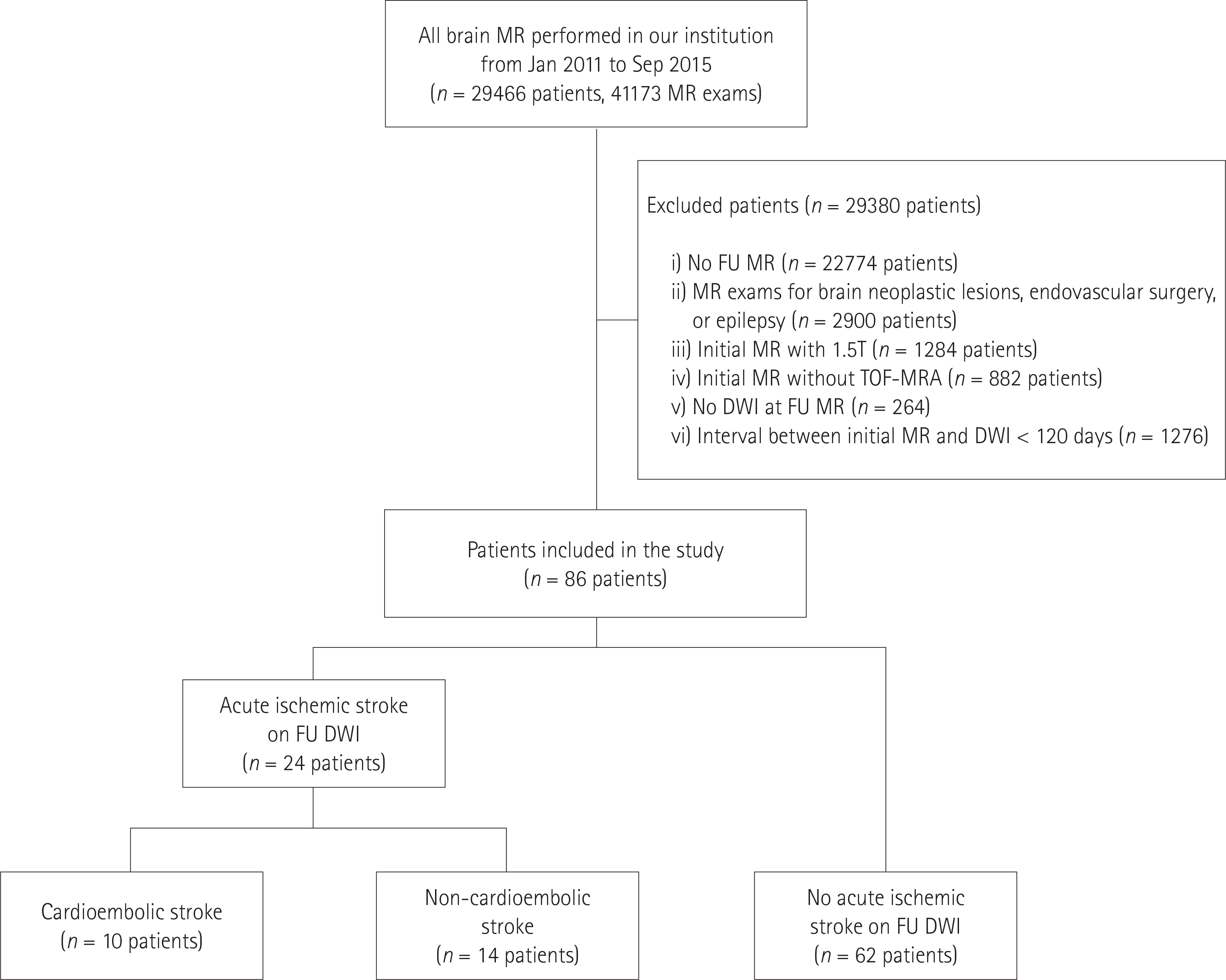
This retrospective study included 86 patients without acute ischemic stroke who first underwent magnetic resonance imaging (MRI) including the time-of-flight magnetic resonance angiography (TOF-MRA) at 3T. Diffusion-weighted imaging (DWI) was performed for the follow-up imaging of these patients > 120 days after the initial MRI. The TOF-MRA result was used to analyze three morphological characteristics: dilatation, stenosis, and tortuosity. The presence of acute ischemic stroke was assessed using the follow-up DWI data. We built two prognostic models: model 1 includes the conventional stroke-risk factors, while model 2 includes the conventional risk factors and the morphologic characteristics of the intracranial arteries. We used the likelihood-ratio test to compare these two models. The models' performances were evaluated using Harrell's concordance index.
RESULTS
Fourteen patients suffered non-cardioembolic strokes. The performances of the two models differed significantly regarding the future-risk modeling of the non-cardioembolic stroke (p = 0.031). The Harrell's concordance index of model 2 (0.78 ± 0.05) exceeded that of model 1 (0.72 ± 0.07).
CONCLUSION
In addition to the conventional stroke-risk factors, the morphologic characteristics of the intracranial arteries were useful in the modeling of the future risk of the non-cardioembolic ischemic stroke.
MeSH Terms
Figure
Reference
-
References
1. Han J, Qiao H, Li X, Li X, He Q, Wang Y, et al. The three-dimensional shape analysis of the M1 segment of the middle cerebral artery using MRA at 3T. Neuroradiology. 2014; 56:995–1005.
Article2. Diedrich KT, Roberts JA, Schmidt RH, Kang CK, Cho ZH, Parker DL. Validation of an arterial tortuosity measure with application to hypertension collection of clinical hypertensive patients. BMC bioinformatics. 2011; 12(Suppl 10):S15.
Article3. Pico F, Labreuche J, Touboul PJ, Amarenco P. Intracranial arterial dolichoectasia and its relation with atherosclerosis and stroke subtype. Neurology. 2003; 61:1736–1742.
Article4. Pico F, Labreuche J, Touboul PJ, Leys D, Amarenco P. Intracranial arterial dolichoectasia and small-vessel disease in stroke patients. Ann Neurol. 2005; 57:472–479.
Article5. Del Corso L, Moruzzo D, Conte B, Agelli M, Romanelli AM, Pastine F, et al. Tortuosity, kinking, and coiling of the carotid artery: expression of atherosclerosis or aging? Angiology. 1998; 49:361–371.
Article6. MacDougall NJ, Amarasinghe S, Muir KW. Secondary prevention of stroke. Expert Rev Cardiovasc Ther. 2009; 7:1103–1115.
Article7. Gutierrez J, Sacco RL, Wright CB. Dolichoectasia-an evolving arterial disease. Nat Rev Neurol. 2011; 7:41–50.
Article8. Çoban G, Çifçi E, Yildirim E, Ag˘ ıldere AM. Predisposing factors in posterior circulation infarcts: a vascular morphological assessment. Neuroradiology. 2015; 57:483–489.
Article9. Nakamura Y, Hirayama T, Ikeda K. Clinicoradiologic features of vertebrobasilar dolichoectasia in stroke patients. J Stroke Cerebrovasc Dis. 2012; 21:5–10.10. Qiao Y, Anwar Z, Intrapiromkul J, Liu L, Zeiler SR, Leigh R, et al. Patterns and implications of intracranial arterial remodeling in stroke patients. Stroke. 2016; 47:434–440.
Article11. Lee WJ, Choi HS, Jang J, Sung J, Kim TW, Koo J, et al. Non-stenotic intracranial arteries have atherosclerotic changes in acute ischemic stroke patients: a 3T MRI study. Neuroradiology. 2015; 57:1007–1013.
Article12. Morris SA, Orbach DB, Geva T, Singh MN, Gauvreau K, Lacro RV. Increased vertebral artery tortuosity index is associated with adverse outcomes in children and young adults with connective tissue disorders. Circulation. 2011; 124:388–396.
Article13. Deguchi I, Ohe Y, Fukuoka T, Dembo T, Nagoya H, Kato Y, et al. Relationship of obesity to recanalization after hyperacute recombinant tissue-plasminogen activator infusion therapy in patients with middle cerebral artery occlusion. J Stroke Cerebrovasc Dis. 2012; 21:161–164.
Article14. Samuels OB, Joseph GJ, Lynn MJ, Smith HA, Chimowitz MI. A standardized method for measuring intracranial arterial stenosis. AJNR Am J Neuroradiol. 2000; 21:643–646.15. Kapeller P, Barber R, Vermeulen RJ, Adèr H, Scheltens P, Fre-idl W, et al. Visual rating of age-related white matter changes on magnetic resonance imaging: scale comparison, interrater agreement, and correlations with quantitative measurements. Stroke. 2003; 34:441–445.16. North American Symptomatic Carotid Endarterectomy Trial Collaborators; Barnett HJM, Taylor DW, Haynes RB, Sackett DL, Peerless SJ, Ferguson GG, et al. Beneficial effect of carotid endarterectomy in symptomatic patients with high-grade carotid stenosis. N Engl J Med. 1991; 325:445–453.
Article17. Arboix A, Garcia-Eroles L, Massons JB, Oliveres M, Pujades R, Targa C. Atrial fibrillation and stroke: clinical presentation of cardioembolic versus atherothrombotic infarction. Int J Cardiol. 2000; 73:33–42.
Article18. Martí-Vilalta JL, Matías-Guiu J. [Nomenclature of cerebral vascular diseases]. Neurologia. 1987; 2:166–175.19. Li ML, Xu WH, Song L, Feng F, You H, Ni J, et al. Atherosclerosis of middle cerebral artery: evaluation with high-resolution MR imaging at 3T. Atherosclerosis. 2009; 204:447–452.
Article20. Degnan AJ, Gallagher G, Teng Z, Lu J, Liu Q, Gillard JH. MR angiography and imaging for the evaluation of middle cerebral artery atherosclerotic disease. AJNR Am J Neuroradiol. 2012; 33:1427–1435.
Article21. Ince B, Petty GW, Brown RD Jr, Chu CP, Sicks JD, Whisnant JP. Dolichoectasia of the intracranial arteries in patients with first ischemic stroke: a population-based study. Neurology. 1998; 50:1694–1698.
Article22. Gillies RJ, Kinahan PE, Hricak H. Radiomics: images are more than pictures, they are data. Radiology. 2016; 278:563–577.
Article23. Adams HP Jr, Bendixen BH, Kappelle LJ, Biller J, Love BB, Gordon DL, et al. Classification of subtype of acute ischemic stroke. definitions for use in a multicenter clinical trial. TOAST. trial of Org 10172 in acute stroke treatment. Stroke. 1993; 24:35–41.
Article24. Arboix A, Alió J. Acute cardioembolic stroke: an update. Expert Rev Cardiovasc Ther. 2011; 9:367–379.
Article25. Galanis T, Merli GJ. Direct thrombin and factor Xa inhibition for stroke prevention in patients with atrial fibrillation. Hosp Pract (1995). 2013; 41:26–36.
Article26. Arboix A, Oliveres M, Massons J, Pujades R, Garcia-Eroles L. Early differentiation of cardioembolic from atherothrombotic cerebral infarction: a multivariate analysis. Eur J Neurol. 1999; 6:677–683.
Article27. Sacco RL, Benjamin EJ, Broderick JP, Dyken M, Easton JD, Feinberg WM, et al. American heart association prevention conference. IV. prevention and rehabilitation of stroke. risk factors. Stroke. 1997; 28:1507–1517.28. Harmsen P, Lappas G, Rosengren A, Wilhelmsen L. Long-term risk factors for stroke: twenty-eight years of follow-up of 7457 middle-aged men in Göteborg, Sweden. Stroke. 2006; 37:1663–1667.29. Wolf PA, D'Agostino RB, Belanger AJ, Kannel WB. Probability of stroke: a risk profile from the Framingham Study. Stroke. 1991; 22:312–318.
Article
- Full Text Links
- Actions
-
Cited
- CITED
-
- Close
- Share
- Similar articles
-
- The Association between Neurological Prognosis and Serum Glucose Level by Stroke Subtype in Acute Ischemic Stroke
- Simultaneous Onset of Ischemic and Hemorrhagic Stroke Due To Intracranial Artery Dissection
- Effects of Gamma-Glutamyl Transferase on Stroke Occurrence Mediated by Atrial Fibrillation
- Pharmacological Secondary Prevention of Ischemic Stroke
- Percutaneous Transluminal Angioplasty of Intracranial Artery for the Treatment of Acute Ischemic Stroke


